The Fibrillarin/U3 RNP Monoclonal Antibody (CAB0850) is a high-quality antibody developed for reliable detection and analysis of target proteins. This gene product is a component of a nucleolar small nuclear ribonucleoprotein (snRNP) particle thought to participate in the first step in processing preribosomal RNA. It is associated with the U3, U8, and U13 small nuclear RNAs and is located in the dense fibrillar component (DFC) of the nucleolus. The encoded protein contains an N-terminal repetitive domain that is rich in glycine and arginine residues, like fibrillarins in other species. Its central region resembles an RNA-binding domain and contains an RNP consensus sequence. Antisera from approximately 8% of humans with the autoimmune disease scleroderma recognize fibrillarin. RRID AB_2861482 Gene ID 2091 Swiss Prot Synonym FIB; FLRN; Nop1; RNU3IP1; Fibrillarin/U3 RNP
This antibody is validated for use in WB, IF/ICC, IP, ELISA applications and has demonstrated reactivity against Human, Mouse, Rat samples.
Product Name:
Fibrillarin/U3 RNP Monoclonal Antibody
SKU:
CAB0850
Size:
100μL, 20μL
Reactivity:
Human, Mouse, Rat
Clone Number:
ARC0506
Conjugate:
Unconjugated
Immunogen:
Synthetic peptide. This information is considered to be commercially sensitive.
Tested Applications:
WBIF/ICCIPELISA
Recommended Dilution:
WB
1:1000 - 1:10000
IF
/
ICC
1:100 - 1:1000
IP
0.5μg-4μg antibody for 200μg-400μg extracts of whole cells
ELISA
Recommended starting concentration is 1 μg/mL. Please optimize the concentration based on your specific assay requirements.
Synonyms:
FIB, FLRN, Nop1, RNU3IP1, Fibrillarin/U3 RNP
Positive Sample:
293T, Hep G2, Jurkat, Mouse spleen, Rat liver, 293F
Cellular Localization:
Nucleus, Nucleolus.
Calculated MW:
34kDa
Observed MW:
37kDa
This gene product is a component of a nucleolar small nuclear ribonucleoprotein (snRNP) particle thought to participate in the first step in processing preribosomal RNA. It is associated with the U3, U8, and U13 small nuclear RNAs and is located in the dense fibrillar component (DFC) of the nucleolus. The encoded protein contains an N-terminal repetitive domain that is rich in glycine and arginine residues, like fibrillarins in other species. Its central region resembles an RNA-binding domain and contains an RNP consensus sequence. Antisera from approximately 8% of humans with the autoimmune disease scleroderma recognize fibrillarin. RRID AB_2861482 Gene ID 2091 Swiss Prot Synonym FIB; FLRN; Nop1; RNU3IP1; Fibrillarin/U3 RNP
Purification Method:
Affinity purification
Gene ID:
2091
RRID:
AB_2861482
Buffer Information:
Store at -20℃. Avoid freeze / thaw cycles. Buffer: PBS containing 50% glycerol and 0.05% BSA, preserved with proclin300 or sodium azide, pH 7.3.
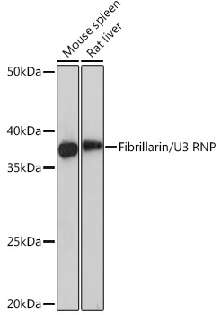
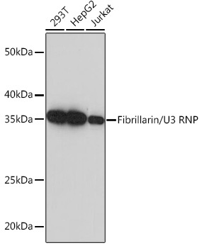
Western blot analysis of various lysates using Fibrillarin/U3 RNP Rabbit mAb (CAB0850) at 1:1000 dilution. Secondary antibody: HRP-conjugated Goat anti-Rabbit IgG (H+L) (AS014) at 1:10000 dilution. Lysates/proteins: 25μg per lane. Blocking buffer: 3% nonfat dry milk in TBST. Detection: ECL Basic Kit (AbGn00020). Exposure time: 10s.
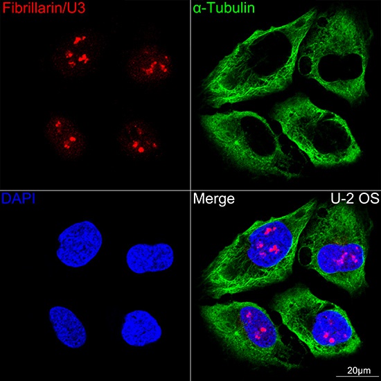
Confocal imaging of U-2OS cells using Fibrillarin/U3 RNP Rabbit mAb (CAB0850,dilution 1:100) (Red) . The cells were counterstained with α-Tubulin Mouse mAb (AC012,dilution 1:400) (Green). DAPI was used for nuclear staining (blue). Objective: 60x.
Confocal imaging of U-2OS cells using Fibrillarin/U3 RNP Rabbit mAb (CAB0850,dilution 1:100) (Red) . The cells were counterstained with α-Tubulin Mouse mAb (AC012,dilution 1:400) (Green). DAPI was used for nuclear staining (blue). Objective: 60x.
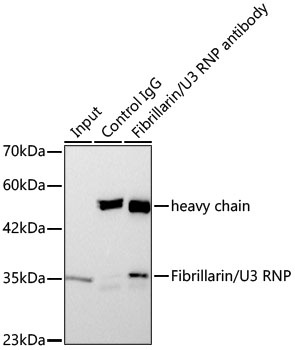
Immunoprecipitation of Fibrillarin/U3 RNP Rabbit mAb from 300 µg extracts of 293F cells was performed using 3 µg of Fibrillarin/U3 RNP Rabbit mAb (CAB0850). Rabbit IgG isotype control (AC042) was used to precipitate the Control IgG sample. IP samples were eluted with 1X Laemmli Buffer. The Input lane represents 10% of the total input. Western blot analysis of immunoprecipitates was conducted using Fibrillarin/U3 RNP Rabbit mAb (CAB0850) at a dilution of 1 : 2000.