The FUBP1 Monoclonal Antibody (CAB9077) is a high-quality antibody developed for reliable detection and analysis of target proteins. The protein encoded by this gene is a single stranded DNA-binding protein that binds to multiple DNA elements, including the far upstream element (FUSE) located upstream of c-myc. Binding to FUSE occurs on the non-coding strand, and is important to the regulation of c-myc in undifferentiated cells. This protein contains three domains, an amphipathic helix N-terminal domain, a DNA-binding central domain, and a C-terminal transactivation domain that contains three tyrosine-rich motifs. The N-terminal domain is thought to repress the activity of the C-terminal domain. This protein is also thought to bind RNA, and contains 3'-5' helicase activity with in vitro activity on both DNA-DNA and RNA-RNA duplexes. Aberrant expression of this gene has been found in malignant tissues, and this gene is important to neural system and lung development. Binding of this protein to viral RNA is thought to play a role in several viral diseases, including hepatitis C and hand, foot and mouth disease. Alternative splicing results in multiple transcript variants.
This antibody is validated for use in IHC-P, IF/ICC, IP, ELISA, IF-P applications and has demonstrated reactivity against Human, Mouse, Rat samples.
Product Name:
FUBP1 Monoclonal Antibody
SKU:
CAB9077
Size:
100μL, 20μL
Reactivity:
Human, Mouse, Rat
Clone Number:
ARC1403
Conjugate:
Unconjugated
Immunogen:
Synthetic peptide. This information is considered to be commercially sensitive.
Tested Applications:
IHC-PIF/ICCIPELISAIF-P
Recommended Dilution:
IP
0.5μg-4μg antibody for 200μg-400μg extracts of whole cells
IF/ICC
1:100 - 1:500
IF-P
1:100 - 1:500
IHC-P
1:50 - 1:200
ELISA
Recommended starting concentration is 1 μg/mL. Please optimize the concentration based on your specific assay requirements.
Synonyms:
FBP, FUBP, hDH V, FUBP1
Positive Sample:
Rat brain,
Cellular Localization:
Cytoplasm, Nucleoplasm, Nnucleus.
Calculated MW:
68kDa
Observed MW:
68kDa/70kDa
The protein encoded by this gene is a single stranded DNA-binding protein that binds to multiple DNA elements, including the far upstream element (FUSE) located upstream of c-myc. Binding to FUSE occurs on the non-coding strand, and is important to the regulation of c-myc in undifferentiated cells. This protein contains three domains, an amphipathic helix N-terminal domain, a DNA-binding central domain, and a C-terminal transactivation domain that contains three tyrosine-rich motifs. The N-terminal domain is thought to repress the activity of the C-terminal domain. This protein is also thought to bind RNA, and contains 3'-5' helicase activity with in vitro activity on both DNA-DNA and RNA-RNA duplexes. Aberrant expression of this gene has been found in malignant tissues, and this gene is important to neural system and lung development. Binding of this protein to viral RNA is thought to play a role in several viral diseases, including hepatitis C and hand, foot and mouth disease. Alternative splicing results in multiple transcript variants.
Purification Method
Affinity purification
Gene ID
8880
Buffer Information
Store at -20℃. Avoid freeze / thaw cycles. Buffer: PBS containing 50% glycerol and 0.05% BSA, preserved with proclin300 or sodium azide, pH 7.3.
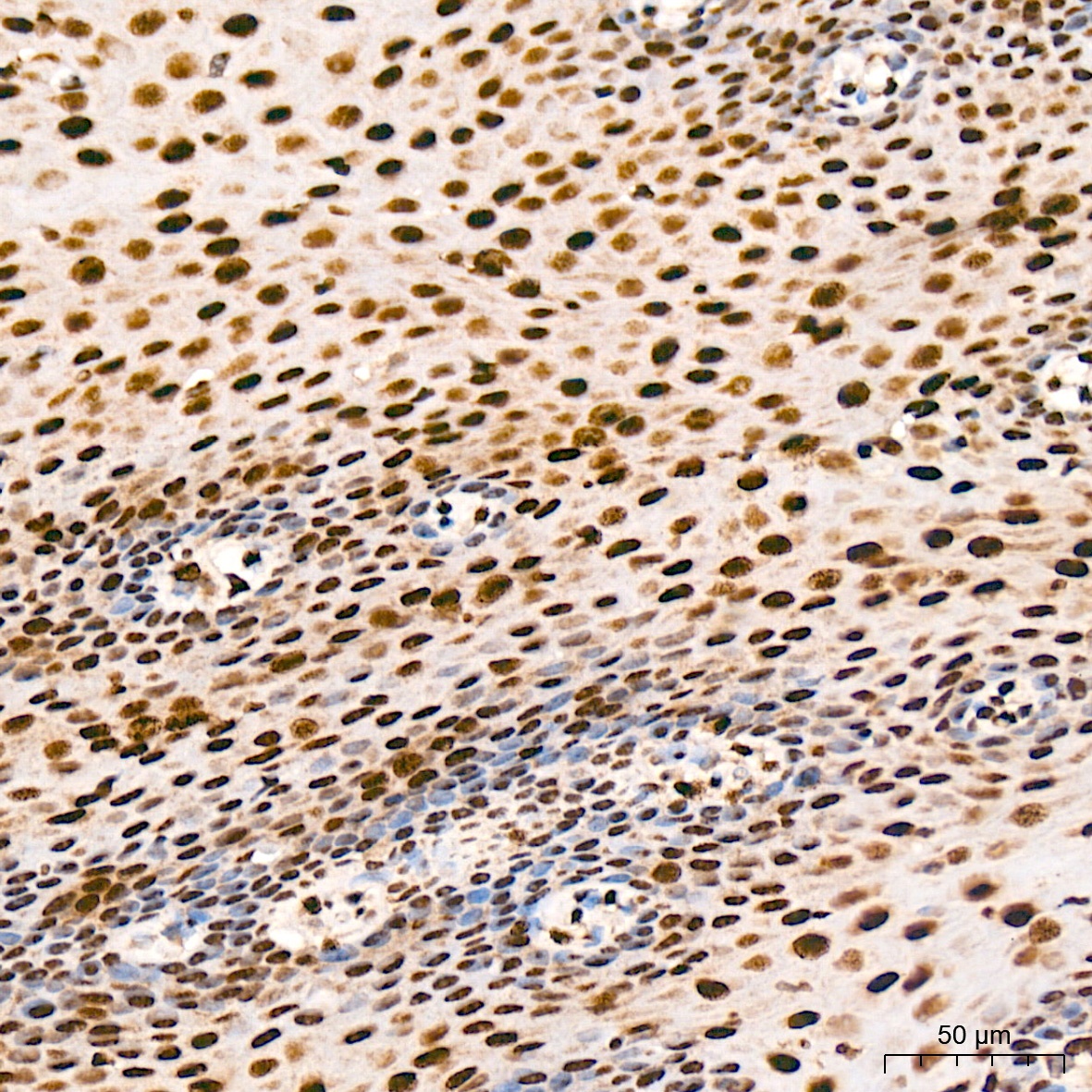
Immunohistochemistry analysis of paraffin-embedded Human thyroid tissue using FUBP1 Rabbit mAb (CAB9077) at a dilution of 1:200 (40x lens). High pressure antigen retrieval was performed with 0.01 M citrate buffer (pH 6.0) prior to IHC staining.
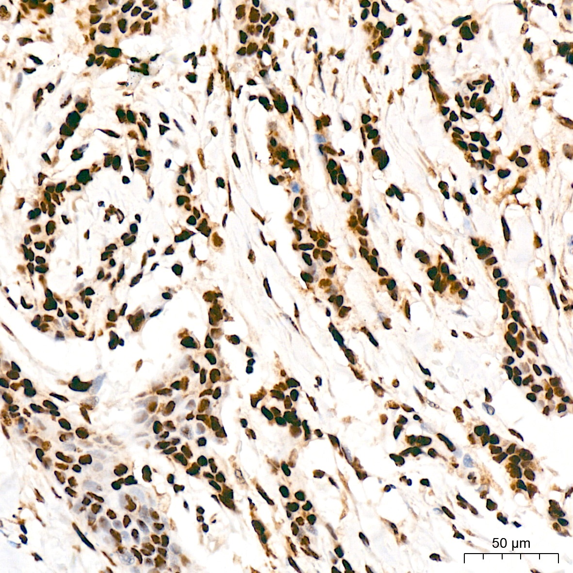
Immunohistochemistry analysis of paraffin-embedded Human breast cancer tissue using FUBP1 Rabbit mAb (CAB9077) at a dilution of 1:200 (40x lens). High pressure antigen retrieval was performed with 0.01 M citrate buffer (pH 6.0) prior to IHC staining.
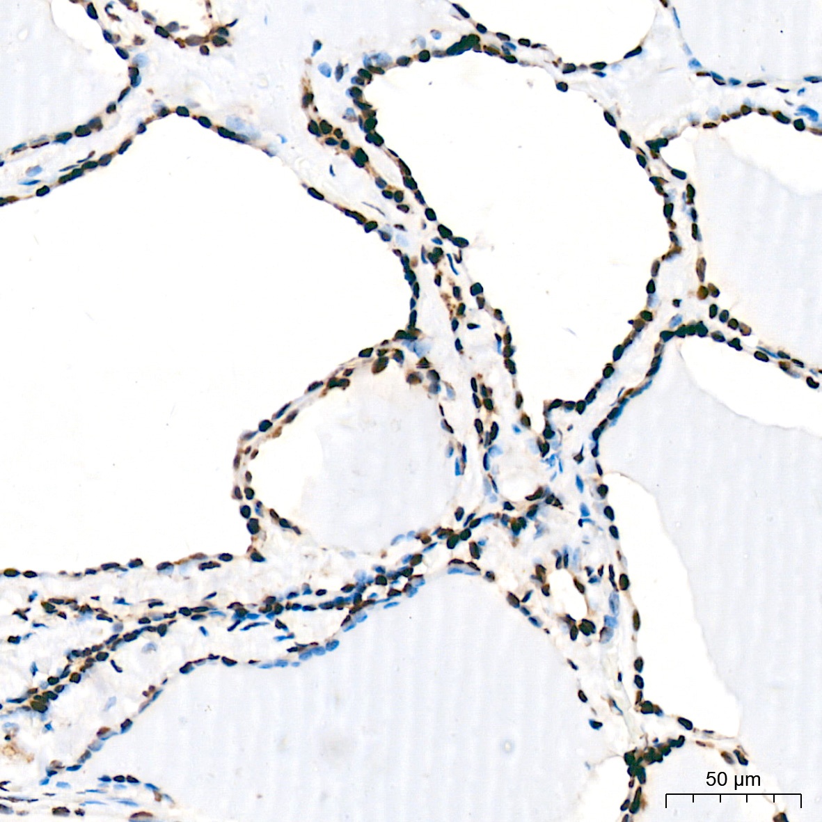
Immunohistochemistry analysis of paraffin-embedded Mouse lung tissue using FUBP1 Rabbit mAb (CAB9077) at a dilution of 1:200 (40x lens). High pressure antigen retrieval was performed with 0.01 M citrate buffer (pH 6.0) prior to IHC staining.
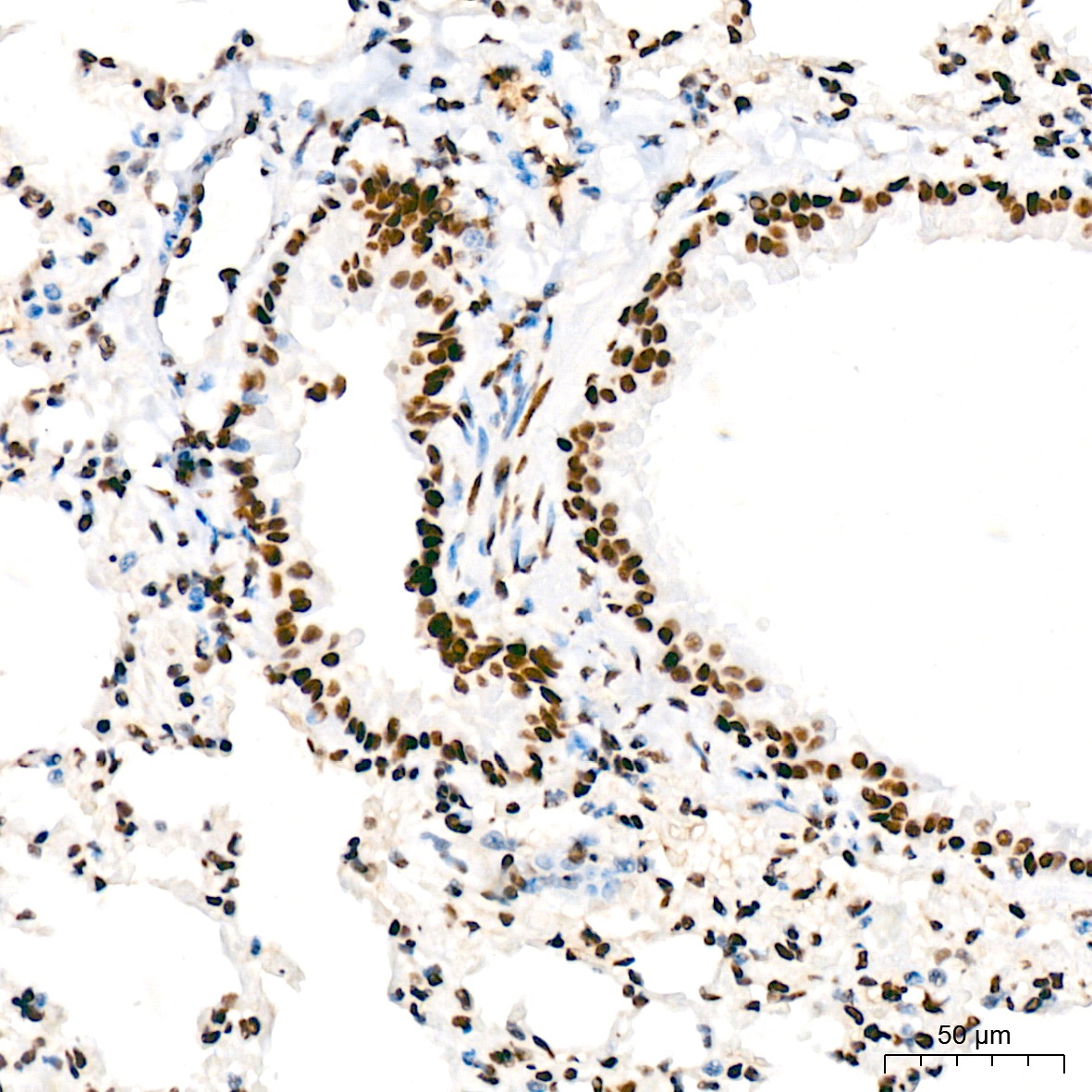
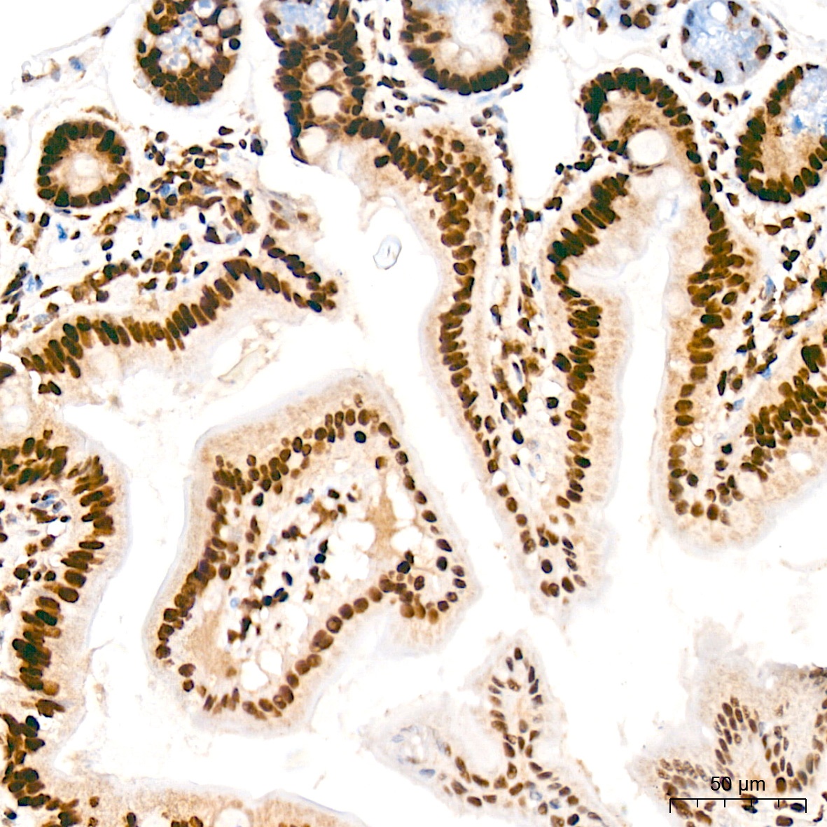
Immunohistochemistry analysis of paraffin-embedded Human esophagus tissue using FUBP1 Rabbit mAb (CAB9077) at a dilution of 1:200 (40x lens). High pressure antigen retrieval was performed with 0.01 M citrate buffer (pH 6.0) prior to IHC staining.
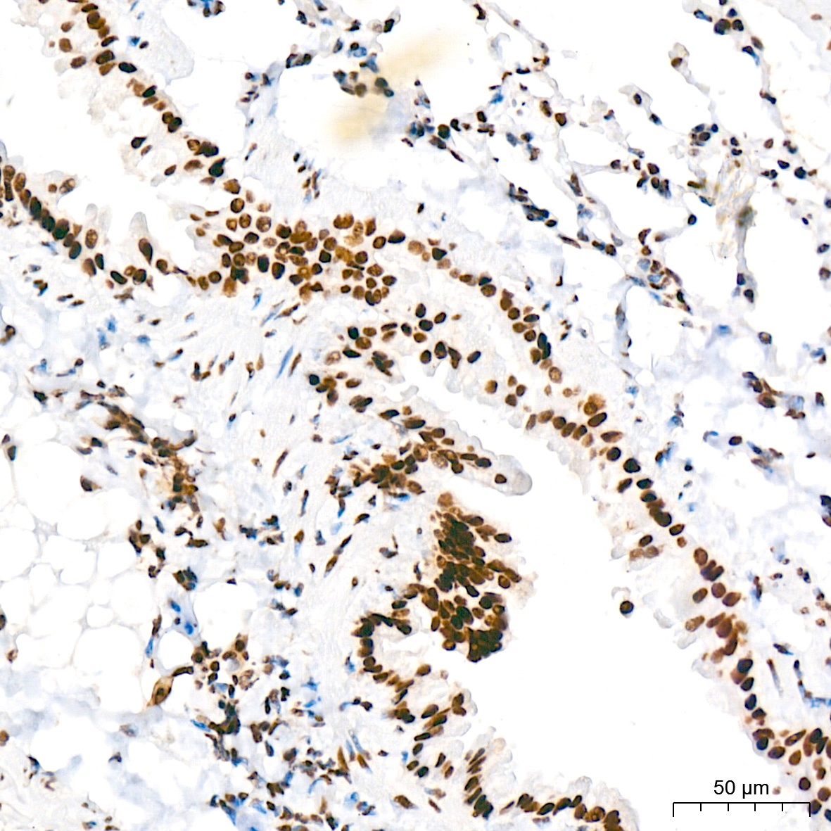
Immunohistochemistry analysis of paraffin-embedded Rat lung tissue using FUBP1 Rabbit mAb (CAB9077) at a dilution of 1:200 (40x lens). High pressure antigen retrieval was performed with 0.01 M citrate buffer (pH 6.0) prior to IHC staining.
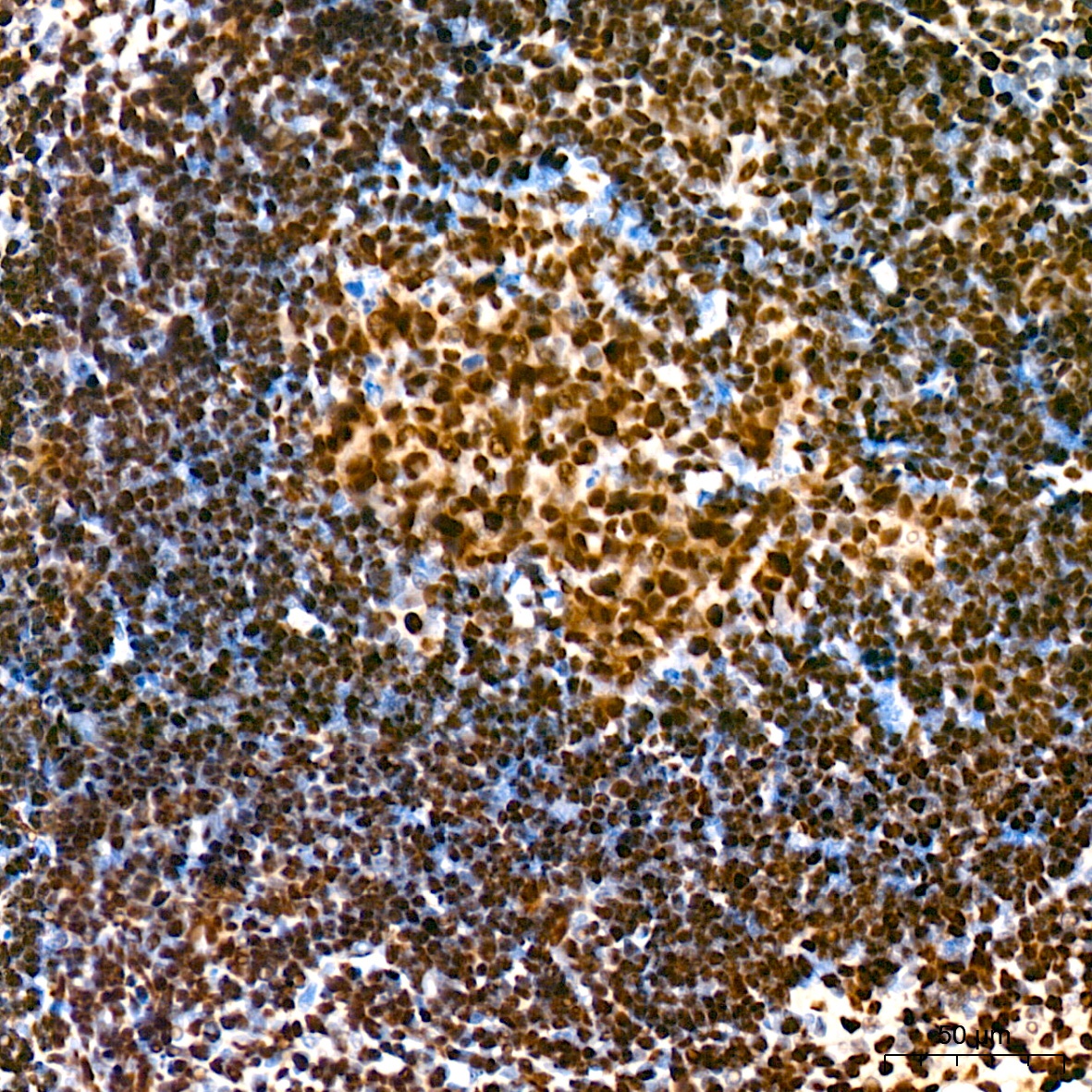
Immunohistochemistry analysis of paraffin-embedded Mouse spleen tissue using FUBP1 Rabbit mAb (CAB9077) at a dilution of 1:200 (40x lens). High pressure antigen retrieval was performed with 0.01 M citrate buffer (pH 6.0) prior to IHC staining.
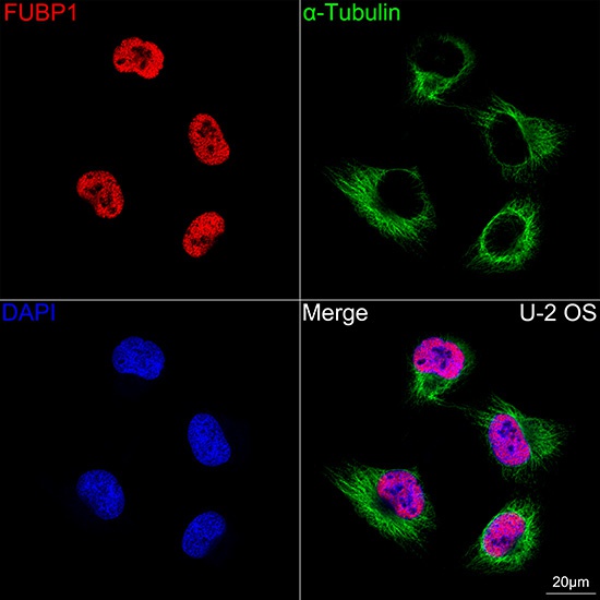
Confocal imaging of U-2 OS cells using FUBP1 Rabbit mAb (CAB9077, dilution 1:300) followed by a further incubation with Cy3 Goat Anti-Rabbit IgG (H+L) (AS007, dilution 1:500) (Red). The cells were counterstained with α-Tubulin Mouse mAb (AC012, dilution 1:400) followed by incubation with ABflo® 488-conjugated Goat Anti-Mouse IgG (H+L) Ab (AS076, dilution 1:500) (Green). DAPI was used for nuclear staining (Blue). Objective: 100x.
Confocal imaging of U-2 OS cells using FUBP1 Rabbit mAb (CAB9077, dilution 1:300) followed by a further incubation with Cy3 Goat Anti-Rabbit IgG (H+L) (AS007, dilution 1:500) (Red). The cells were counterstained with α-Tubulin Mouse mAb (AC012, dilution 1:400) followed by incubation with ABflo® 488-conjugated Goat Anti-Mouse IgG (H+L) Ab (AS076, dilution 1:500) (Green). DAPI was used for nuclear staining (Blue). Objective: 100x.
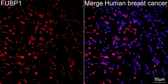
Confocal imaging of paraffin-embedded Human breast cancer tissue using FUBP1 Rabbit mAb (CAB9077, dilution 1:300) followed by a further incubation with Cy3 Goat Anti-Rabbit IgG (H+L) (AS007, dilution 1:500) (Red). DAPI was used for nuclear staining (Blue). Objective: 40x. Perform high pressure antigen retrieval with 0.01 M citrate buffer (pH 6.0) prior to IF staining.
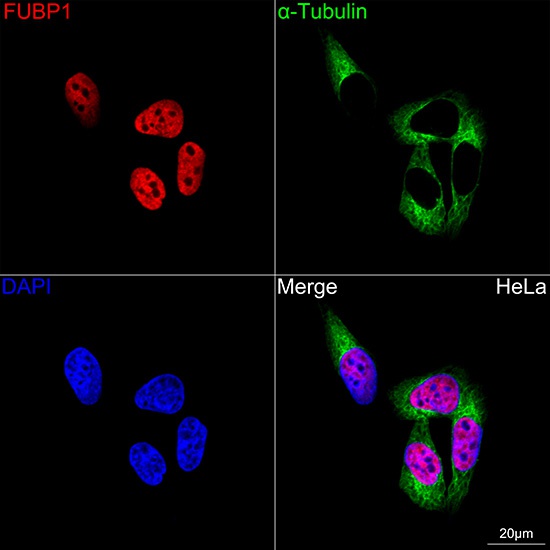
Confocal imaging of HeLa cells using FUBP1 Rabbit mAb (CAB9077, dilution 1:300) followed by a further incubation with Cy3 Goat Anti-Rabbit IgG (H+L) (AS007, dilution 1:500) (Red). The cells were counterstained with α-Tubulin Mouse mAb (AC012, dilution 1:400) followed by incubation with ABflo® 488-conjugated Goat Anti-Mouse IgG (H+L) Ab (AS076, dilution 1:500) (Green). DAPI was used for nuclear staining (Blue). Objective: 100x.