The HCoV-229E Spike S1 Antibody (CAB20393) is a high-quality antibody developed for reliable detection and analysis of target proteins. This antibody, generated in rabbits, specifically targets the Spike S1 protein of the virus, which plays a key role in viral attachment and entry into host cells.Validated for use in various applications such as ELISA, immunofluorescence, and Western blot, this antibody allows for the detection and analysis of the HCoV-229E Spike S1 protein in human samples. Its high reactivity and specificity make it an invaluable resource for studying the molecular mechanisms of HCoV-229E infection, as well as for developing diagnostic tools and potential therapeutic interventions.
This antibody is validated for use in WB, ELISA applications and has demonstrated reactivity against HCoV-229E samples.
Product Name:
HCoV-229E Spike S1 Antibody
SKU:
CAB20393
Size:
20μL, 100μL
Reactivity:
HCoV-229E
Conjugate:
Unconjugated
Immunogen:
Synthetic peptide. This information is considered to be commercially sensitive.
Recommended starting concentration is 1 μg/mL. Please optimize the concentration based on your specific assay requirements.
Positive Sample:
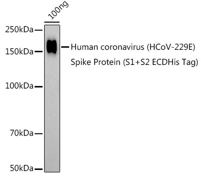
Human coronavirus (HCoV-229E) Spike Protein (S1+S2 ECDHis Tag)
Calculated MW:
129kDa
Observed MW:
160kDa
S1 region attaches the virion to the cell membrane by interacting with host ANPEP/aminopeptidase N, initiating the infection. Binding to the receptor probably induces conformational changes in the S glycoprotein unmasking the fusion peptide of S2 region and activating membranes fusion. S2 region belongs to the class I viral fusion protein. Under the current model, the protein has at least 3 conformational states: pre-fusion native state, pre-hairpin intermediate state, and post-fusion hairpin state. During viral and target cell membrane fusion, the coiled coil regions (heptad repeats regions assume a trimer-of-hairpins structure, positioning the fusion peptide in close proximity to the C-terminal region of the ectodomain. The formation of this structure appears to drive apposition and subsequent fusion of viral and target cell membranes.
Purification Method
Affinity purification
Gene ID
918758
Buffer Information
Store at -20℃. Avoid freeze / thaw cycles. Buffer: PBS containing 50% glycerol, preserved with proclin300 or sodium azide, pH 7.3.
Western blot analysis of lysates from Human coronavirus (HCoV-229E) Spike Protein (S1+S2 ECDHis Tag), using HCoV-229E Spike S1 Rabbit pAb (CAB20393) at 1:1000 dilution. Secondary antibody: HRP-conjugated Goat anti-Rabbit IgG (H+L) (CABS014) at 1:10000 dilution. Lysates/proteins: 25μg per lane. Blocking buffer: 3% nonfat dry milk in TBST. Detection: ECL Basic Kit (AbGn00020). Exposure time: 180s.