LI Cadherin/Cadherin-17 Monoclonal Antibody (CAB5286)
The LI Cadherin/Cadherin-17 Monoclonal Antibody (CAB5286) is a high-quality antibody developed for reliable detection and analysis of target proteins. This antibody, produced in rabbits, is highly specific for LI-Cadherin and has been validated for use in various applications, including Western blotting and immunohistochemistry.LI-Cadherin, also known as Cadherin-17, is predominantly expressed in the gastrointestinal tract, where it is involved in maintaining the integrity of the epithelial barrier and regulating cell proliferation and differentiation.
This antibody is validated for use in WB, IHC-P, ELISA, IF-P applications and has demonstrated reactivity against Human, Mouse, Rat samples.
Product Name:
LI Cadherin/Cadherin-17 Monoclonal Antibody
SKU:
CAB5286
Size:
20μL, 100μL
Reactivity:
Human, Mouse, Rat
Clone Number:
ARC1989
Conjugate:
Unconjugated
Immunogen:
Synthetic peptide. This information is considered to be commercially sensitive.
Recommended starting concentration is 1 μg/mL. Please optimize the concentration based on your specific assay requirements.
Synonyms:
HPT1, CDH16, HPT-1, LI Cadherin/Cadherin-17
Positive Sample:
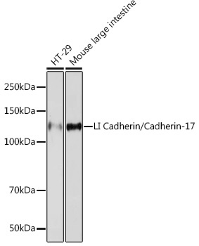
HT-29 cells, Mouse large intestine
Cellular Localization:
Cell Membrane, Single-Pass Type I Membrane Protein.
Calculated MW:
92kDa
Observed MW:
120kDa
This gene is a member of the cadherin superfamily, genes encoding calcium-dependent, membrane-associated glycoproteins. The encoded protein is cadherin-like, consisting of an extracellular region, containing 7 cadherin domains, and a transmembrane region but lacking the conserved cytoplasmic domain. The protein is a component of the gastrointestinal tract and pancreatic ducts, acting as an intestinal proton-dependent peptide transporter in the first step in oral absorption of many medically important peptide-based drugs. The protein may also play a role in the morphological organization of liver and intestine. Alternative splicing results in multiple transcript variants.
Purification Method
Affinity purification
Gene ID
1015
RRID
AB_2863498
Buffer Information
Store at -20℃. Avoid freeze / thaw cycles. Buffer: PBS containing 50% glycerol and 0.05% BSA, preserved with proclin300 or sodium azide, pH 7.3.
Western blot analysis of various lysates using LI Cadherin/Cadherin-17 Rabbit mAb (CAB5286) at 1:1000 dilution. Secondary antibody: HRP-conjugated Goat anti-Rabbit IgG (H+L) (CABS014) at 1:10000 dilution. Lysates/proteins: 25μg per lane. Blocking buffer: 3% nonfat dry milk in TBST. Detection: ECL Basic Kit (AbGn00020). Exposure time: 90s.
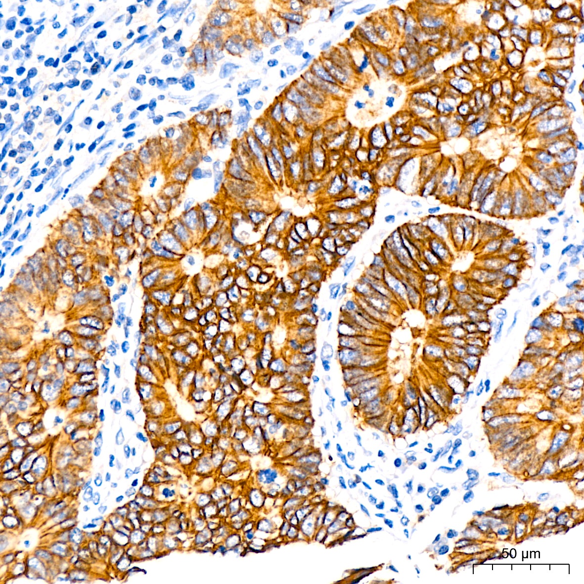
Immunohistochemistry analysis of paraffin-embedded Human gastric cancer using LI Cadherin/Cadherin-17 Rabbit mAb (CAB5286) at dilution of 1:200 (40x lens). High pressure antigen retrieval performed with 0.01M Tris/EDTA Buffer (pH 9.0) prior to IHC staining.
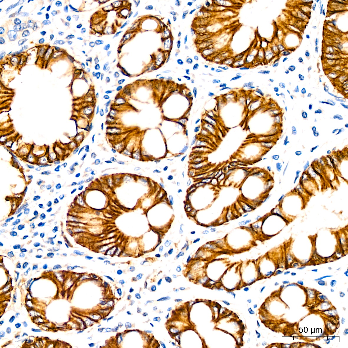
Immunohistochemistry analysis of paraffin-embedded Human stomach using LI Cadherin/Cadherin-17 Rabbit mAb (CAB5286) at dilution of 1:200 (40x lens). High pressure antigen retrieval performed with 0.01M Tris/EDTA Buffer (pH 9.0) prior to IHC staining.
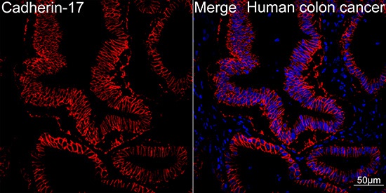
Confocal imaging of Human colon cancer using LI Cadherin/Cadherin-17 Rabbit mAb (CAB5286,dilution 1:100)(Red). DAPI was used for nuclear staining (blue). Objective: 40x.
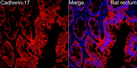
Immunofluorescence analysis of paraffin-embedded rat rectum using LI Cadherin/Cadherin-17 Rabbit mAb (CAB5286) at dilution of 1:100 (40x lens). Secondary antibody: Cy3-conjugated Goat anti-Rabbit IgG (H+L) (CABS007) at 1:500 dilution. Blue: DAPI for nuclear staining.