The MonoMethyl-Histone H3-K18 Monoclonal Antibody (CAB20680) is a high-quality antibody developed for reliable detection and analysis of target proteins. This antibody specifically targets the monomethylated form of histone H3 at lysine 18, a post-translational modification associated with gene activation and transcriptional regulation.Raised in rabbits, this antibody is highly specific and sensitive when detecting monomethyl histone H3 (K18) in human samples. It has been validated for use in Western blot applications, allowing for accurate analysis of histone modifications in different cell types and biological contexts.Histone modifications, such as monomethylation of histone H3 at K18, play a key role in gene expression regulation and epigenetic control.
This antibody is validated for use in WB, IHC-P, IF/ICC, IP, ChIP, ELISA, DB, CUT&Tag applications and has demonstrated reactivity against Human, Mouse, Rat, Other (Wide Range Predicted) samples.
Product Name:
MonoMethyl-Histone H3-K18 Monoclonal Antibody
SKU:
CAB20680
Size:
20μL, 100μL
Reactivity:
Human, Mouse, Rat, Other (Wide Range Predicted)
Clone Number:
ARC2621
Conjugate:
Unconjugated
Immunogen:
Synthetic peptide. This information is considered to be commercially sensitive.
Sequence:
APRK Q
Tested Applications:
WBIHC-PIF/ICCIPChIPELISADBCUT&Tag
Recommended Dilution:
WB
1:500 - 1:1000
DB
1:500 - 1:1000
IHC-P
1:50 - 1:200
IF/ICC
1:50 - 1:200
IP
2μg-6μg antibody for 400μg-600μg extracts of whole cells
ChIP
5μg antibody for 5μg-10μg of Chromatin
CUT&Tag
10⁵ cells /1 μg
ELISA
Recommended starting concentration is 1 μg/mL. Please optimize the concentration based on your specific assay requirements.
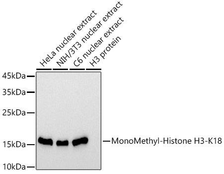
HeLa nuclear extract, NIH/3T3 nuclear extract, C6 nuclear extract
Cellular Localization:
Chromosome, Nucleus.
Calculated MW:
15kDa
Observed MW:
17kDa
Histones are basic nuclear proteins that are responsible for the nucleosome structure of the chromosomal fiber in eukaryotes. Nucleosomes consist of approximately 146 bp of DNA wrapped around a histone octamer composed of pairs of each of the four core histones (H2A, H2B, H3, and H4). The chromatin fiber is further compacted through the interaction of a linker histone, H1, with the DNA between the nucleosomes to form higher order chromatin structures. This gene is intronless and encodes a replication-dependent histone that is a member of the histone H3 family. Transcripts from this gene lack polyA tails; instead, they contain a palindromic termination element. This gene is located separately from the other H3 genes that are in the histone gene cluster on chromosome 6p22-p21.3.
Purification Method
Affinity purification
Gene ID
8290 8350
Buffer Information
Store at -20℃. Avoid freeze / thaw cycles. Buffer: PBS containing 50% glycerol and 0.05% BSA, preserved with proclin300 or sodium azide, pH 7.3.
Western blot analysis of various lysates using MonoMethyl-Histone H3-K18 Rabbit mAb (CAB20680) at 1:1000 dilution. Secondary antibody: HRP-conjugated Goat anti-Rabbit IgG (H+L) (CABS014) at 1:10000 dilution. Lysates/proteins: 25μg per lane. Blocking buffer: 3% nonfat dry milk in TBST. Detection: ECL Basic Kit (AbGn00020). Exposure time: 30s.
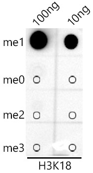
Dot-blot analysis of all sorts of peptides using MonoMethyl-Histone H3-K18 antibody (CAB20680) at 1:1000 dilution.
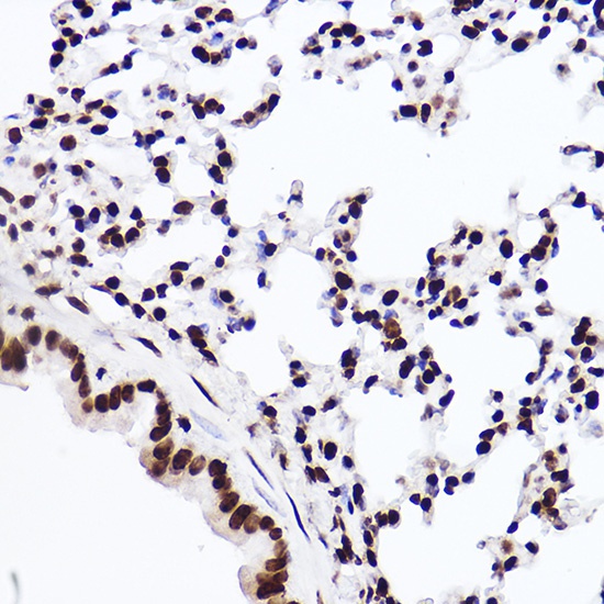
Immunohistochemistry analysis of paraffin-embedded Mouse lung using MonoMethyl-Histone H3-K18 Rabbit mAb (CAB20680) at dilution of 1:100 (40x lens). High pressure antigen retrieval performed with 0.01M Citrate buffer (pH 6.0) prior to IHC staining.
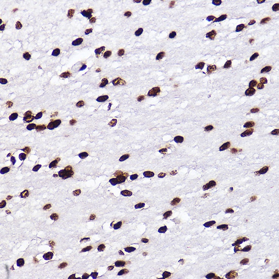
Immunohistochemistry analysis of paraffin-embedded Rat brain using MonoMethyl-Histone H3-K18 Rabbit mAb (CAB20680) at dilution of 1:100 (40x lens). High pressure antigen retrieval performed with 0.01M Citrate buffer (pH 6.0) prior to IHC staining.
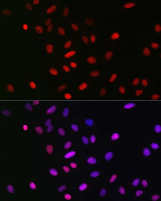
Immunofluorescence analysis of U-2 OS cells using MonoMethyl-Histone H3-K18 Rabbit mAb (CAB20680) at dilution of 1:100 (40x lens). Secondary antibody: Cy3-conjugated Goat anti-Rabbit IgG (H+L) (CABS007) at 1:500 dilution. Blue: DAPI for nuclear staining.
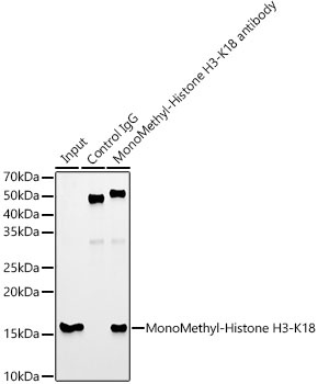
Immunoprecipitation analysis of 600 μg extracts of 293F cells using 5 μg MonoMethyl-Histone H3-K18 antibody (CAB20680). Western blot was performed from the immunoprecipitate using MonoMethyl-Histone H3-K18 antibody (CAB20680) at a dilution of 1:1000.
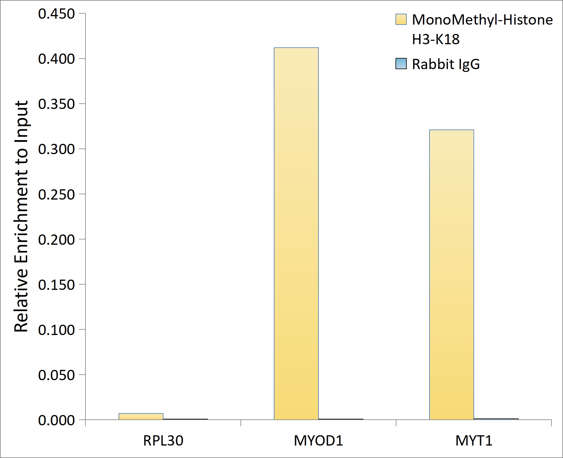
Chromatin immunoprecipitation analysis of extracts of HeLa cells, using MonoMethyl-Histone H3-K18 antibody (CAB20680) and rabbit IgG.The amount of immunoprecipitated DNA was checked by quantitative PCR. Histogram was constructed by the ratios of the immunoprecipitated DNA to the input.
. Blue: DAPI for nuclear staining.")

. Blue: DAPI for nuclear staining.")
. Perform high pressure antigen retrieval with 10 mM citrate buffer pH 6. 0 before commencing with IHC staining protocol.")
. Perform high pressure antigen retrieval with 10 mM citrate buffer pH 6. 0 before commencing with IHC staining protocol.")
. Perform high pressure antigen retrieval with 10 mM citrate buffer pH 6. 0 before commencing with IHC staining protocol.")
 at 1:10000 dilution. Lysates/proteins: 25ug per lane. Blocking buffer: 3% nonfat dry milk in TBST. Detection: ECL Basic Kit. Exposure time: 30s.")
. Blue: DAPI for nuclear staining.")


 at 1:10000 dilution. Lysates/proteins: 25ug per lane. Blocking buffer: 3% nonfat dry milk in TBST. Detection: ECL Basic Kit. Exposure time: 10s.")

