The mSin3A Monoclonal Antibody (CAB2396) is a high-quality antibody developed for reliable detection and analysis of target proteins. This antibody, produced in rabbits, offers high reactivity with human samples and has been validated for use in Western blot applications. By targeting the MSIN3A protein, this antibody allows for the detection and analysis of MSIN3A in various cell types, making it ideal for investigations in molecular biology and cancer research.
This antibody is validated for use in IHC-P, IF/ICC, ELISA applications and has demonstrated reactivity against Human, Mouse, Rat samples.
Product Name:
mSin3A Monoclonal Antibody
SKU:
CAB2396
Size:
20μL, 100μL
Reactivity:
Human, Mouse, Rat
Clone Number:
ARC0745
Conjugate:
Unconjugated
Immunogen:
Synthetic peptide. This information is considered to be commercially sensitive.
The protein encoded by this gene is a transcriptional regulatory protein. It contains paired amphipathic helix (PAH) domains, which are important for protein-protein interactions and may mediate repression by the Mad-Max complex.
Purification Method
Affinity purification
Gene ID
25942
Buffer Information
Store at -20℃. Avoid freeze / thaw cycles. Buffer: PBS containing 50% glycerol and 0.05% BSA, preserved with proclin300 or sodium azide, pH 7.3.
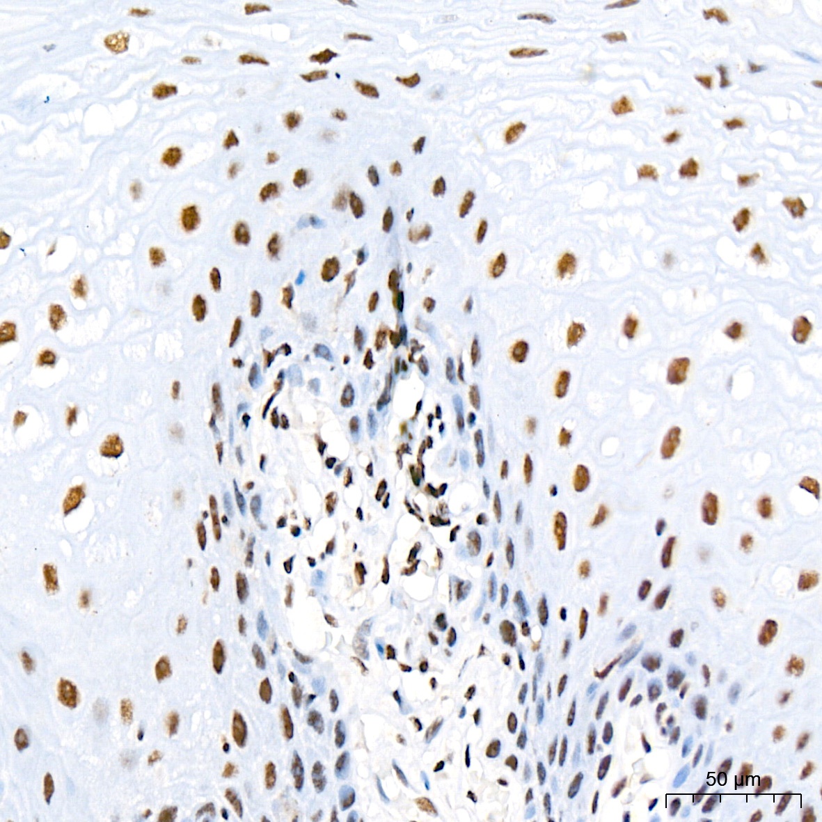
Immunohistochemistry analysis of paraffin-embedded Human esophagus tissue using mSin3A Rabbit mAb (CAB2396) at a dilution of 1:200 (40x lens). High pressure antigen retrieval was performed with 0.01 M citrate buffer (pH 6.0) prior to IHC staining.
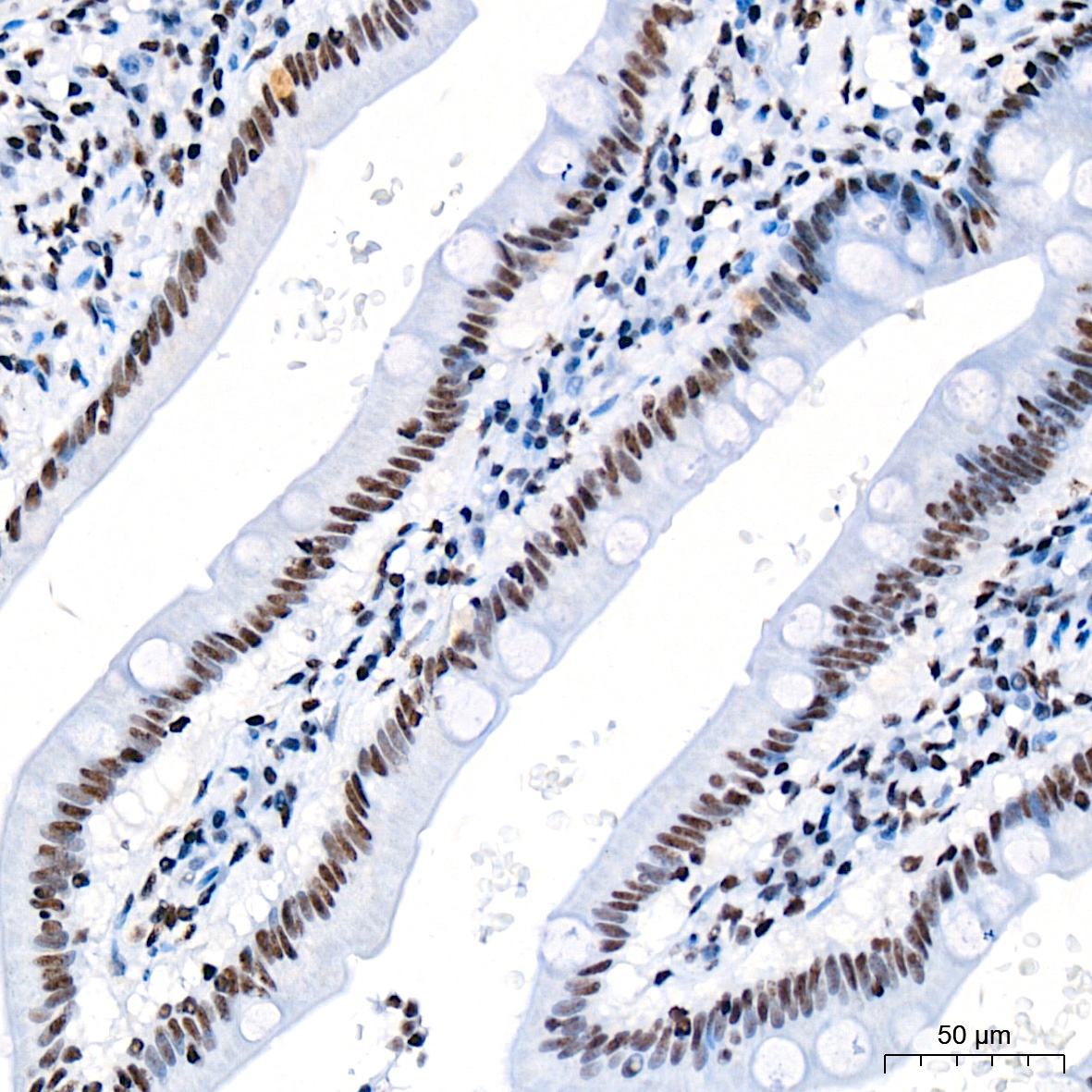
Immunohistochemistry analysis of paraffin-embedded Human small intestine tissue using mSin3A Rabbit mAb (CAB2396) at a dilution of 1:200 (40x lens). High pressure antigen retrieval was performed with 0.01 M citrate buffer (pH 6.0) prior to IHC staining.
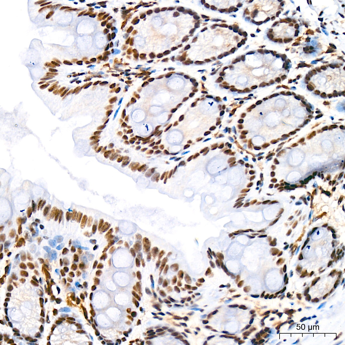
Immunohistochemistry analysis of paraffin-embedded Mouse colon tissue using mSin3A Rabbit mAb (CAB2396) at a dilution of 1:200 (40x lens). High pressure antigen retrieval was performed with 0.01 M citrate buffer (pH 6.0) prior to IHC staining.
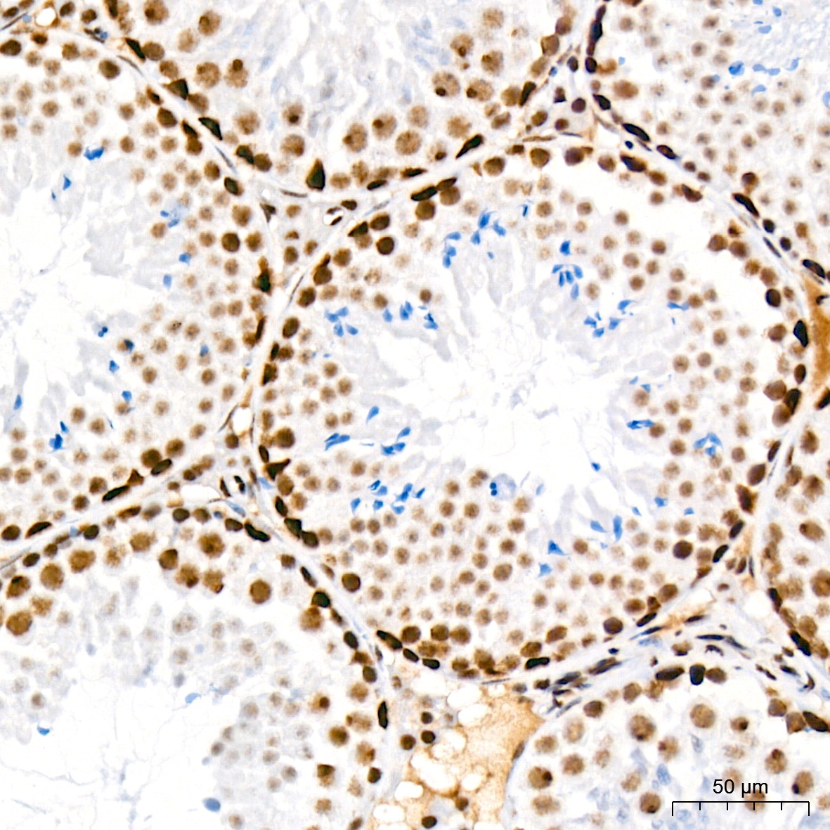
Immunohistochemistry analysis of paraffin-embedded Mouse testis tissue using mSin3A Rabbit mAb (CAB2396) at a dilution of 1:200 (40x lens). High pressure antigen retrieval was performed with 0.01 M citrate buffer (pH 6.0) prior to IHC staining.
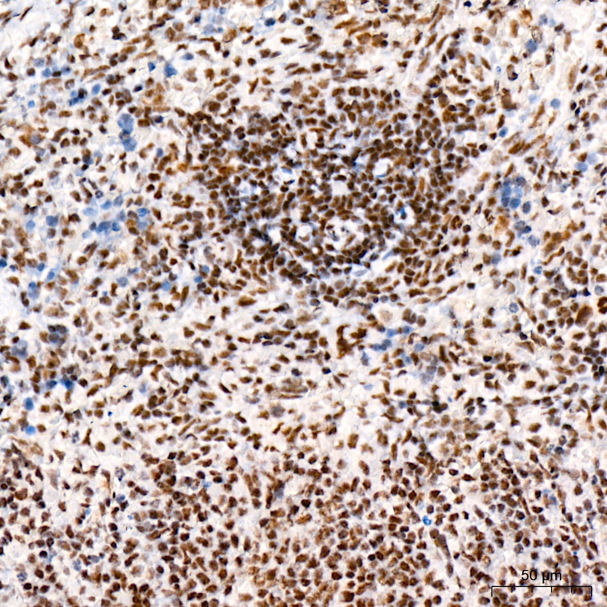
Immunohistochemistry analysis of paraffin-embedded Rat spleen tissue using mSin3A Rabbit mAb (CAB2396) at a dilution of 1:200 (40x lens). High pressure antigen retrieval was performed with 0.01 M citrate buffer (pH 6.0) prior to IHC staining.
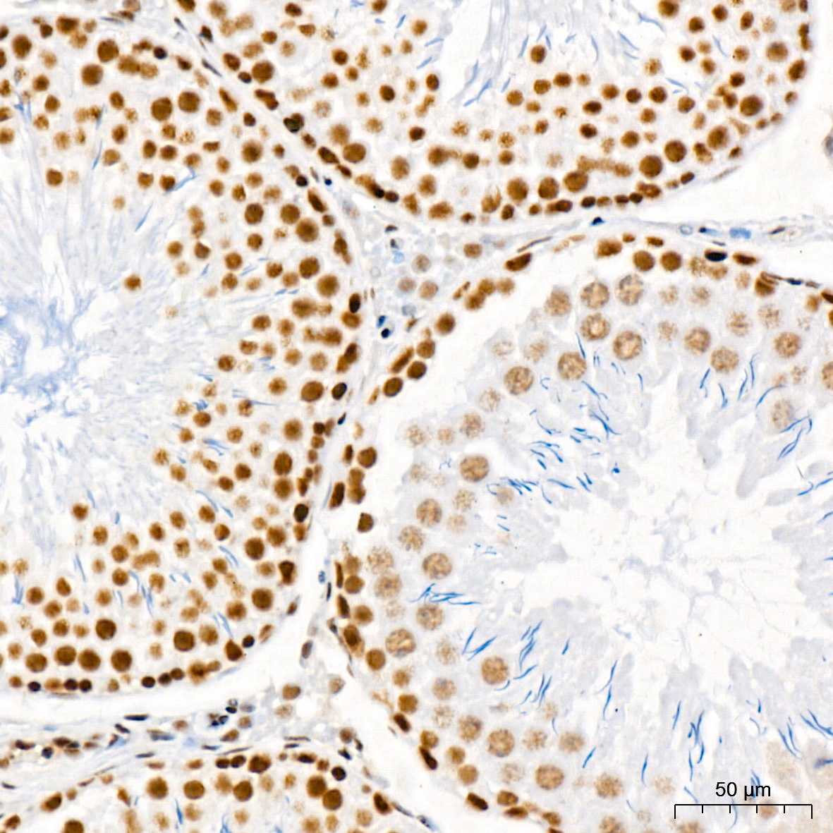
Immunohistochemistry analysis of paraffin-embedded Rat testis tissue using mSin3A Rabbit mAb (CAB2396) at a dilution of 1:200 (40x lens). High pressure antigen retrieval was performed with 0.01 M citrate buffer (pH 6.0) prior to IHC staining.
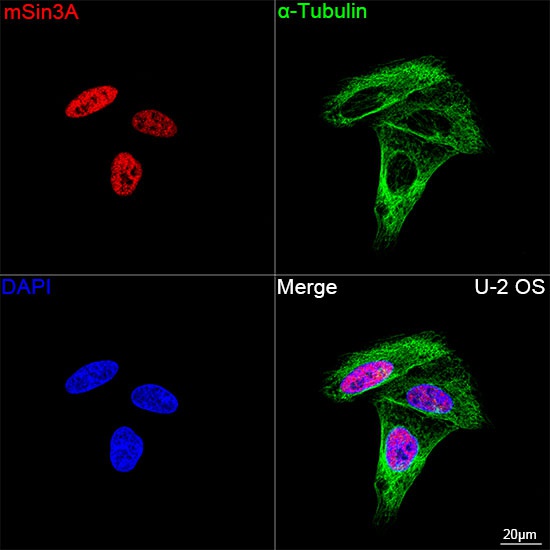
Confocal imaging of U-2 OS cells using mSin3A Rabbit mAb (CAB2396, dilution 1:200) followed by a further incubation with Cy3 Goat Anti-Rabbit IgG (H+L) (CABS007, dilution 1:500) (Red). The cells were counterstained with α-Tubulin Mouse mAb (AC012, dilution 1:400) followed by incubation with ABflo® 488-conjugated Goat Anti-Mouse IgG (H+L) Ab (CABS076, dilution 1:500) (Green). DAPI was used for nuclear staining (Blue). Objective: 100x.