The Musashi-2 (MSI2) Monoclonal Antibody (CAB19814) is a high-quality antibody developed for reliable detection and analysis of target proteins. This gene encodes an RNA-binding protein that is a member of the Musashi protein family. The encoded protein is transcriptional regulator that targets genes involved in development and cell cycle regulation. Mutations in this gene are associated with poor prognosis in certain types of cancers. This gene has also been shown to be rearranged in certain cancer cells.
This antibody is validated for use in WB, IHC-P, ELISA applications and has demonstrated reactivity against Human, Mouse, Rat samples.
Product Name:
Musashi-2 (MSI2) Monoclonal Antibody
SKU:
CAB19814
Size:
100μL, 20μL
Reactivity:
Human, Mouse, Rat
Clone Number:
ARC2341
Conjugate:
Unconjugated
Immunogen:
Synthetic peptide. This information is considered to be commercially sensitive.
Tested Applications:
WBIHC-PELISA
Recommended Dilution:
WB
1:1000 - 1:6000
IHC-P
1:50 - 1:200
ELISA
Recommended starting concentration is 1 μg/mL. Please optimize the concentration based on your specific assay requirements.
Synonyms:
MSI2H, Musashi-2 (MSI2)
Positive Sample:
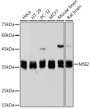
HeLa, HT-29, PC-12, MCF7, Mouse brain, Rat brain
Cellular Localization:
Cytoplasm, Cytosol.
Calculated MW:
35kDa
Observed MW:
35kDa
This gene encodes an RNA-binding protein that is a member of the Musashi protein family. The encoded protein is transcriptional regulator that targets genes involved in development and cell cycle regulation. Mutations in this gene are associated with poor prognosis in certain types of cancers. This gene has also been shown to be rearranged in certain cancer cells.
Purification Method
Affinity purification
Gene ID
124540
Buffer Information
Store at -20℃. Avoid freeze / thaw cycles. Buffer: PBS containing 50% glycerol and 0.05% BSA, preserved with proclin300 or sodium azide, pH 7.3.
Western blot analysis of various lysates using Musashi-2 (MSI2) Rabbit mAb (CAB19814) at 1:1000 dilution. Secondary antibody: HRP-conjugated Goat anti-Rabbit IgG (H+L) (AS014) at 1:10000 dilution. Lysates/proteins: 25μg per lane. Blocking buffer: 3% nonfat dry milk in TBST. Detection: ECL Basic Kit (AbGn00020). Exposure time: 5s.
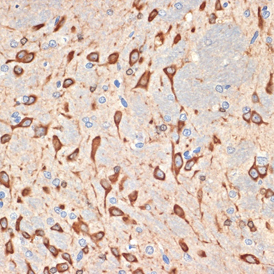
Immunohistochemistry analysis of paraffin-embedded Rat brain tissue using Musashi-2 (MSI2) Rabbit mAb (CAB19814) at dilution of 1:100 (40x lens). Microwave antigen retrieval performed with 0.01M Tris/EDTA Buffer (pH 9.0) prior to IHC staining.