The Neurofilament L Monoclonal Antibody (CAB20269) is a high-quality antibody developed for reliable detection and analysis of target proteins. This antibody, produced in rabbits, demonstrates high reactivity with human samples and has been validated for use in various applications, including immunohistochemistry and Western blotting.Neurofilament L, a major intermediate filament protein in neurons, is essential for axonal growth and maintenance. Abnormalities in neurofilament L expression have been implicated in neurodegenerative diseases such as Alzheimer's and ALS, making it a promising target for diagnostic and therapeutic research.
This antibody is validated for use in WB, IHC-P, ELISA, IF-P applications and has demonstrated reactivity against Human, Mouse, Rat samples.
Product Name:
Neurofilament L Monoclonal Antibody
SKU:
CAB20269
Size:
20μL, 100μL
Reactivity:
Human, Mouse, Rat
Clone Number:
ARC50056
Conjugate:
Unconjugated
Immunogen:
Recombinant protein (or fragment).This information is considered to be commercially sensitive.
Neurofilaments are type IV intermediate filament heteropolymers composed of light, medium, and heavy chains. Neurofilaments comprise the axoskeleton and they functionally maintain the neuronal caliber. They may also play a role in intracellular transport to axons and dendrites. This gene encodes the light chain neurofilament protein. Mutations in this gene cause Charcot-Marie-Tooth disease types 1F (CMT1F) and 2E (CMT2E), disorders of the peripheral nervous system that are characterized by distinct neuropathies. A pseudogene has been identified on chromosome Y.
Purification Method
Affinity purification
Gene ID
4747
Buffer Information
Store at -20℃. Avoid freeze / thaw cycles. Buffer: PBS containing 50% glycerol and 0.05% BSA, preserved with proclin300 or sodium azide, pH 7.3
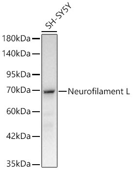
Western blot analysis of lysates from SH-SY5Y cells using Neurofilament L Rabbit mAb (CAB20269) at 1:100000 dilution. Secondary antibody: HRP-conjugated Goat anti-Rabbit IgG (H+L) (CABS014) at 1:10000 dilution. Lysates/proteins: 25 μg per lane. Blocking buffer: 3% nonfat dry milk in TBST. Detection: ECL Basic Kit (AbGn00020). Exposure time:30s.
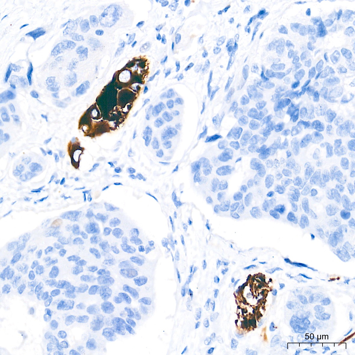
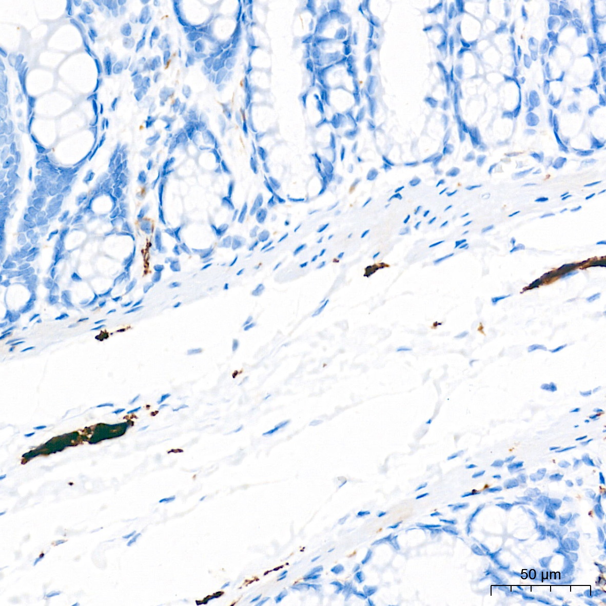
Immunohistochemistry analysis of paraffin-embedded Human colon carcinoma tissue using Neurofilament L Rabbit mAb (CAB20269) at a dilution of 1:1000 (40x lens). High pressure antigen retrieval performed with 0.01M Tris-EDTA Buffer (pH 9.0) prior to IHC staining.
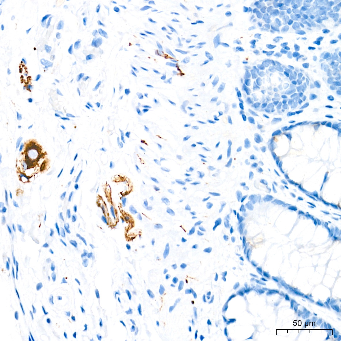
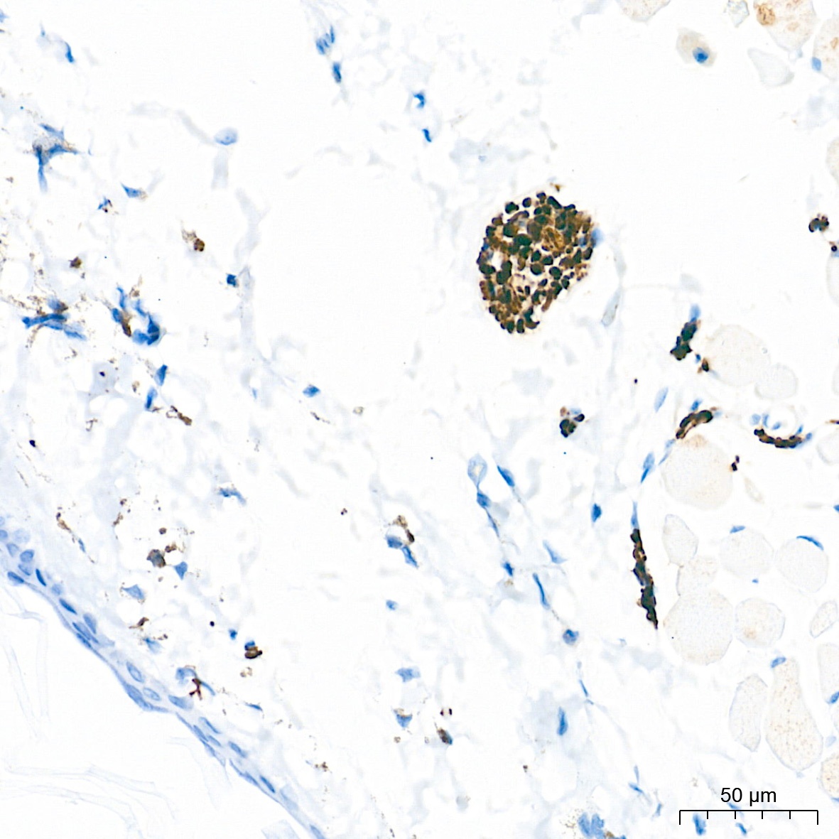
Immunohistochemistry analysis of paraffin-embedded Human colon tissue using Neurofilament L Rabbit mAb (CAB20269) at a dilution of 1:1000 (40x lens). High pressure antigen retrieval performed with 0.01M Tris-EDTA Buffer (pH 9.0) prior to IHC staining.
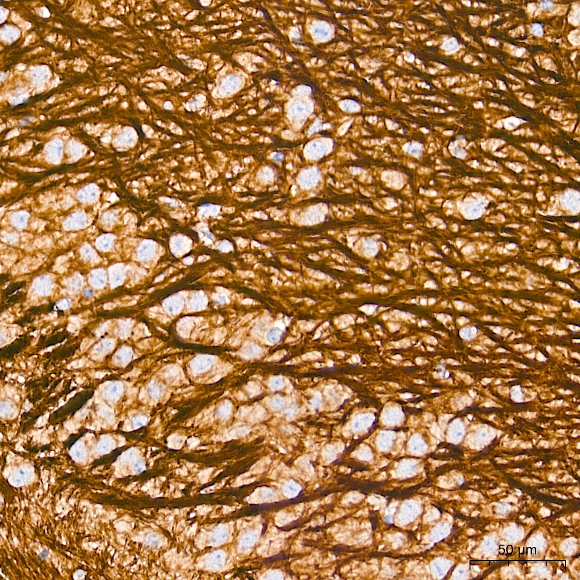
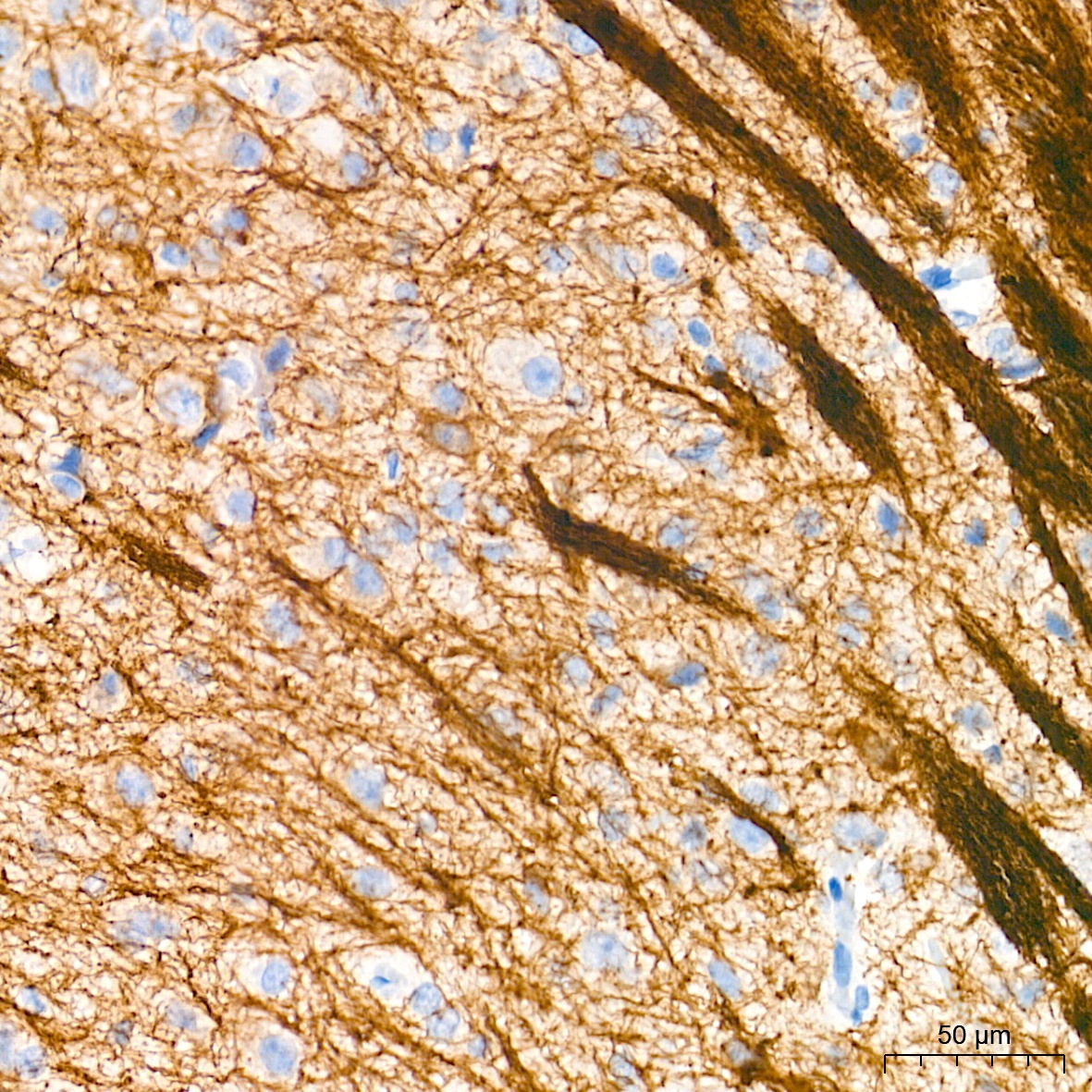
Immunohistochemistry analysis of paraffin-embedded Mouse brain tissue using Neurofilament L Rabbit mAb (CAB20269) at a dilution of 1:1000 (40x lens). High pressure antigen retrieval performed with 0.01M Tris-EDTA Buffer (pH 9.0) prior to IHC staining.
Immunohistochemistry analysis of paraffin-embedded Mouse skin tissue using Neurofilament L Rabbit mAb (CAB20269) at a dilution of 1:1000 (40x lens). High pressure antigen retrieval performed with 0.01M Tris-EDTA Buffer (pH 9.0) prior to IHC staining.
Immunohistochemistry analysis of paraffin-embedded Rat brain tissue using Neurofilament L Rabbit mAb (CAB20269) at a dilution of 1:1000 (40x lens). High pressure antigen retrieval performed with 0.01M Tris-EDTA Buffer (pH 9.0) prior to IHC staining.
Immunohistochemistry analysis of paraffin-embedded Rat colon tissue using Neurofilament L Rabbit mAb (CAB20269) at a dilution of 1:1000 (40x lens). High pressure antigen retrieval performed with 0.01M Tris-EDTA Buffer (pH 9.0) prior to IHC staining.
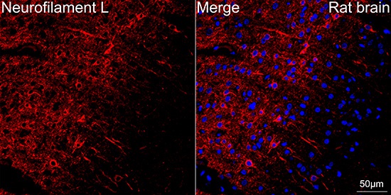
Confocal imaging of paraffin-embedded Rat brain tissue using Neurofilament L Rabbit mAb (CAB20269, dilution 1:200) followed by a further incubation with Cy3 Goat Anti-Rabbit IgG (H+L) (CABS007,dilution 1:500) (Red). DAPI was used for nuclear staining (Blue). Objective: 40x. Perform microwave antigen retrieval with 0.01 M citrate buffer (pH 6.0) prior to IF staining.
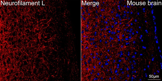
Confocal imaging of paraffin-embedded Mouse brain tissue using Neurofilament L Rabbit mAb (CAB20269, dilution 1:200) followed by a further incubation with Cy3 Goat Anti-Rabbit IgG (H+L) (CABS007, dilution 1:500) (Red). DAPI was used for nuclear staining (Blue). Objective: 40x. Perform microwave antigen retrieval with 0.01 M citrate buffer (pH 6.0) prior to IF staining.