The PFDN5 Monoclonal Antibody (CAB4101) is a high-quality antibody developed for reliable detection and analysis of target proteins. This polyclonal antibody, developed in rabbits, exhibits strong reactivity with human samples and is validated for use in Western blot applications. By binding to the PFDN5 protein, this antibody enables precise detection and analysis in a variety of cell types, making it an indispensable asset for investigations in molecular biology and cellular physiology.PFDN5, also known as prefoldin subunit 5, plays a crucial role in protein folding processes, assisting in the maintenance of protein structure and function.
This antibody is validated for use in WB, IF/ICC, ELISA, IF-P applications and has demonstrated reactivity against Human, Mouse, Rat samples.
Product Name:
PFDN5 Monoclonal Antibody
SKU:
CAB4101
Size:
20μL, 100μL
Reactivity:
Human, Mouse, Rat
Clone Number:
ARC2111
Conjugate:
Unconjugated
Immunogen:
Synthetic peptide. This information is considered to be commercially sensitive.
Sequence:
LNVL NKSN EGKE LLVP LTSS MYVP GKLH DVEH VLID VGTG YYVE KTAE DAKD FFKR KIDF LTKQ MEKI QPAL QEKH AMKQ AVME MMSQ KIQQ LTAL GAAQ ATAK A
Tested Applications:
WBIF/ICCELISAIF-P
Recommended Dilution:
WB
1:500 - 1:1000
IF/ICC
1:50 - 1:200
IF-P
1:50 - 1:200
ELISA
Recommended starting concentration is 1 μg/mL. Please optimize the concentration based on your specific assay requirements.
This gene encodes a member of the prefoldin alpha subunit family. The encoded protein is one of six subunits of prefoldin, a molecular chaperone complex that binds and stabilizes newly synthesized polypeptides, thereby allowing them to fold correctly. The complex, consisting of two alpha and four beta subunits, forms a double beta barrel assembly with six protruding coiled-coils. The encoded protein may also repress the transcriptional activity of the proto-oncogene c-Myc. Alternatively spliced transcript variants encoding different isoforms have been described.
Purification Method
Affinity purification
Gene ID
5204
Buffer Information
Store at -20℃. Avoid freeze / thaw cycles. Buffer: PBS containing 50% glycerol and 0.05% BSA, preserved with proclin300 or sodium azide, pH 7.3.
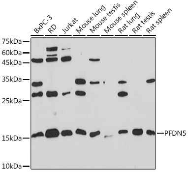
Western blot analysis of various lysates using PFDN5 Rabbit mAb (CAB4101) at 1:1000 dilution. Secondary antibody: HRP-conjugated Goat anti-Rabbit IgG (H+L) (CABS014) at 1:10000 dilution. Lysates/proteins: 25μg per lane. Blocking buffer: 3% nonfat dry milk in TBST. Detection: ECL Basic Kit (AbGn00020). Exposure time: 180s.
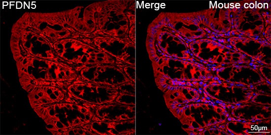
Confocal imaging of paraffin-embedded Mouse colon tissue using PFDN5 Rabbit mAb (CAB4101, dilution 1:100) followed by a further incubation with Cy3 Goat Anti-Rabbit IgG (H+L) (CABS007, dilution 1:500) (Red). DAPI was used for nuclear staining (Blue). Objective: 40x. Perform high pressure antigen retrieval with 0.01 M citrate buffer (pH 6.0) prior to IF staining.