The Phospho-c-Jun-S63 Antibody (CABP1190) is a high-quality antibody developed for reliable detection and analysis of target proteins. This antibody, raised in rabbits, is highly specific for detecting phosphorylated c-Jun protein at serine 63 in human samples. It has been validated for use in Western blot applications, allowing for accurate detection and analysis of phospho c-Jun levels in various cell types.The phosphorylation of c-Jun at serine 63 is known to play a key role in regulating gene expression and cell proliferation, making this antibody a critical tool for researchers studying the molecular mechanisms underlying cancer and inflammatory diseases.
This antibody is validated for use in WB, IHC-P, ELISA applications and has demonstrated reactivity against Human, Mouse, Rat samples.
Product Name:
Phospho-c-Jun-S63 Antibody
SKU:
CABP1190
Size:
20μL, 100μL
Reactivity:
Human, Mouse, Rat
Conjugate:
Unconjugated
Immunogen:
Synthetic peptide. This information is considered to be commercially sensitive.
Sequence:
LTSP D
Tested Applications:
WBIHC-PELISA
Recommended Dilution:
WB
1:500 - 1:1000
IHC-P
1:50 - 1:200
ELISA
Recommended starting concentration is 1 μg/mL. Please optimize the concentration based on your specific assay requirements.
Synonyms:
AP1, p39, AP-1, cJUN, c-Jun, Phospho-c-Jun-S63
Positive Sample:
293T treated with Anisomycin
Cellular Localization:
Nucleus.
Calculated MW:
36kDa
Observed MW:
44kDa
This gene is the putative transforming gene of avian sarcoma virus 17. It encodes a protein which is highly similar to the viral protein, and which interacts directly with specific target DNA sequences to regulate gene expression. This gene is intronless and is mapped to 1p32-p31, a chromosomal region involved in both translocations and deletions in human malignancies.
Purification Method
Affinity purification
Gene ID
3725
RRID
AB_2864046
Buffer Information
Store at -20℃. Avoid freeze / thaw cycles. Buffer: PBS containing 50% glycerol, preserved with proclin300 or sodium azide, pH 7.3.
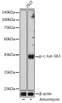
Western blot analysis of lysates from 293T cells, using Phospho-c-Jun-S63 Rabbit pAb (CABP1190) at 1:1000 dilution. 293T cells were treated with Anisomycin (25 μg/mL) at 37℃ for 30 minutes after serum-starvation overnight. Secondary antibody: HRP-conjugated Goat anti-Rabbit IgG (H+L) (CABS014) at 1:10000 dilution. Lysates/proteins: 25μg per lane. Blocking buffer: 3% nonfat dry milk in TBST. Detection: ECL Basic Kit (AbGn00020). Exposure time: 30s.
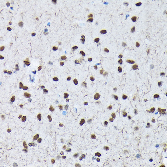
Immunohistochemistry analysis of paraffin-embedded Rat brain using Phospho-c-Jun-S63 Rabbit pAb (CABP1190) at dilution of 1:200 (40x lens). High pressure antigen retrieval performed with 0.01M Citrate buffer (pH 6.0) prior to IHC staining.
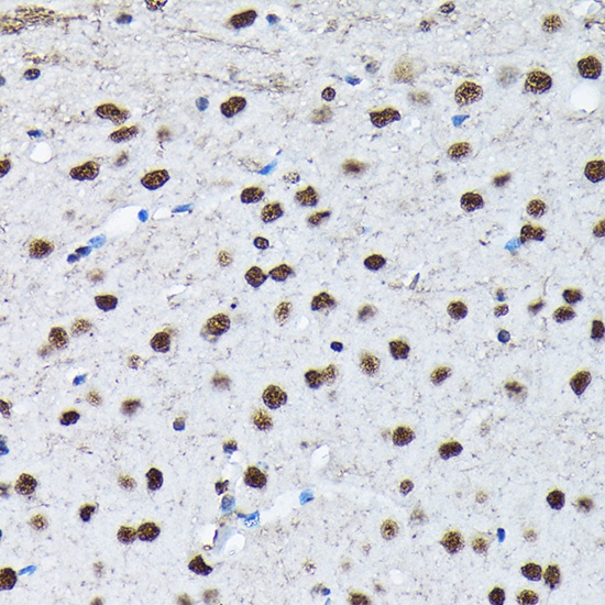
Immunohistochemistry analysis of paraffin-embedded Mouse brain using Phospho-c-Jun-S63 Rabbit pAb (CABP1190) at dilution of 1:200 (40x lens). High pressure antigen retrieval performed with 0.01M Citrate buffer (pH 6.0) prior to IHC staining.