The Phospho-PAK1/2/3-S144/S141/S139 Monoclonal Antibody (CABP1158) is a high-quality antibody developed for reliable detection and analysis of target proteins. This gene encodes a family member of serine/threonine p21-activating kinases, known as PAK proteins. These proteins are critical effectors that link RhoGTPases to cytoskeleton reorganization and nuclear signaling, and they serve as targets for the small GTP binding proteins Cdc42 and Rac. This specific family member regulates cell motility and morphology. Alternatively spliced transcript variants encoding different isoforms have been found for this gene. [provided by RefSeq, Apr 2010]
This antibody is validated for use in WB, ELISA applications and has demonstrated reactivity against Human, Mouse, Rat samples.
Synthetic peptide. This information is considered to be commercially sensitive.
Tested Applications:
WBELISA
Recommended Dilution:
WB
1:500 - 1:1000
ELISA
Recommended starting concentration is 1 μg/mL. Please optimize the concentration based on your specific assay requirements.
Synonyms:
PAKalpha, Phospho-PAK1/2/3-S144/S141/S139
Positive Sample:
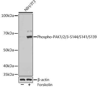
NIH/3T3 treated with Forskolin
Cellular Localization:
Cytoplasm, Cytosol, Focal Adhesion, Microtubule Organizing Center, Nuclear Membrane, Nucleoplasm, Plasma Membrane, Ruffle Membrane, Z Disc.
Calculated MW:
65kDa
Observed MW:
65kDa
This gene encodes a family member of serine/threonine p21-activating kinases, known as PAK proteins. These proteins are critical effectors that link RhoGTPases to cytoskeleton reorganization and nuclear signaling, and they serve as targets for the small GTP binding proteins Cdc42 and Rac. This specific family member regulates cell motility and morphology. Alternatively spliced transcript variants encoding different isoforms have been found for this gene. [provided by RefSeq, Apr 2010]
Purification Method
Affinity purification
Gene ID
5058 5062 5063
RRID
AB_2864020
Buffer Information
Store at -20℃. Avoid freeze / thaw cycles. Buffer: PBS containing 50% glycerol and 0.05% BSA, preserved with proclin300 or sodium azide, pH 7.3.
Western blot analysis of various lysates using Phospho-PAK1/2/3-S144/S141/S139 Rabbit mAb (CABP1158) at 1:1000 dilution. NIH/3T3 cells were treated with Forskolin (30 uM) at 37℃ for 30 minutes after serum-starvation overnight Secondary antibody: HRP-conjugated Goat anti-Rabbit IgG (H+L) (AS014) at 1:10000 dilution. Lysates/proteins: 25μg per lane. Blocking buffer: 3% BSA. Detection: ECL Basic Kit (AbGn00020). Exposure time: 1s.