The Phospho-PKR/EIF2AK2-T446 Antibody (CABP1251) is a high-quality antibody developed for reliable detection and analysis of target proteins. PKR is known for its role in the cellular response to viral infection, stress, and inflammation, and its phosphorylation at T446 is critical for its activity in modulating translation and immune response.This highly specific antibody, raised in rabbits, is validated for use in Western blot applications and has been shown to be highly reactive with human samples. By targeting the phosphorylated form of PKR at T446, researchers can gain insights into the signaling pathways and regulatory mechanisms involving this protein, particularly in the context of viral infections, autoimmune disorders, and cancer.
This antibody is validated for use in WB, IF/ICC, ELISA applications and has demonstrated reactivity against Human, Mouse samples.
Product Name:
Phospho-PKR/EIF2AK2-T446 Antibody
SKU:
CABP1251
Size:
20μL, 100μL
Reactivity:
Human, Mouse
Conjugate:
Unconjugated
Immunogen:
Synthetic peptide. This information is considered to be commercially sensitive.
Sequence:
KRTR SK
Tested Applications:
WBIF/ICCELISA
Recommended Dilution:
WB
1:1000 - 1:5000
IF/ICC
1:50 - 1:200
ELISA
Recommended starting concentration is 1 μg/mL. Please optimize the concentration based on your specific assay requirements.
The protein encoded by this gene is a serine/threonine protein kinase that is activated by autophosphorylation after binding to dsRNA. The activated form of the encoded protein can phosphorylate translation initiation factor EIF2S1, which in turn inhibits protein synthesis. This protein is also activated by manganese ions and heparin. The encoded protein plays an important role in the innate immune response against multiple DNA and RNA viruses.
Purification Method
Affinity purification
Gene ID
5610
Buffer Information
Store at -20℃. Avoid freeze / thaw cycles. Buffer: PBS containing 50% glycerol, preserved with proclin300 or sodium azide, pH 7.3.
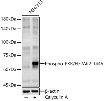
Western blot analysis of lysates from NIH/3T3 cells using Phospho-PKR/EIF2AK2-T446 Rabbit pAb (CABP1251) at 1:2000 dilution. NIH/3T3 cells were treated with Calyculin A (100 nM) at 37℃ for 30 minutes after serum-starvation overnight. Secondary antibody: HRP-conjugated Goat anti-Rabbit IgG (H+L) (CABS014) at 1:10000 dilution. Lysates/proteins: 25 μg per lane. Blocking buffer: 3% nonfat dry milk in TBST. Detection: ECL Basic Kit (AbGn00020). Exposure time: 180s.
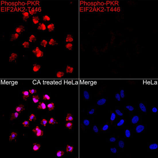
Immunofluorescence analysis of HeLa treated with CA and HeLa cells using Phospho-PKR/EIF2AK2-T446 Rabbit pAb (CABP1251) at dilution of 1:100 (40x lens). Secondary antibody: Cy3-conjugated Goat anti-Rabbit IgG (H+L) (CABS007) at 1:500 dilution. Blue: DAPI for nuclear staining.