The Phospho-RPS6KA5-T581 Polyclonal Antibody (CABP1197) is a high-quality antibody developed for reliable detection and analysis of target proteins. This antibody, generated in rabbits, specifically targets the phosphorylated form of RPS6KA5 at threonine 581 and is highly reactive with human samples. It has been validated for use in Western blot applications, allowing for the detection and analysis of phosphorylated RPS6KA5 in various cell types.RPS6KA5, also known as RSK2, is a serine/threonine kinase with multiple functions in cell signaling and gene expression. Phosphorylation of RPS6KA5 at threonine 581 has been implicated in the regulation of its kinase activity and downstream signaling cascades.
This antibody is validated for use in WB, ELISA applications and has demonstrated reactivity against Rat samples.
Product Name:
Phospho-RPS6KA5-T581 Polyclonal Antibody
SKU:
CABP1197
Size:
20μL, 100μL
Reactivity:
Rat
Conjugate:
Unconjugated
Immunogen:
Synthetic peptide. This information is considered to be commercially sensitive.
Sequence:
Email for sequence
Tested Applications:
WBELISA
Recommended Dilution:
WB
1:500 - 1:1000
ELISA
Recommended starting concentration is 1 μg/mL. Please optimize the concentration based on your specific assay requirements.
Synonyms:
MSK1, RLPK, MSPK1, Phospho-RPS6KA5-T581
Positive Sample:
C6 treated with Calyculin A
Cellular Localization:
Cytoplasm, Nucleus.
Calculated MW:
90kDa
Observed MW:
90kDa
Enables ATP binding activity and protein serine/threonine kinase activity. Involved in several processes, including histone-serine phosphorylation; positive regulation of histone modification; and regulation of transcription, DNA-templated. Located in cytoplasm and nucleoplasm.
Purification Method
Affinity purification
Gene ID
9252
RRID
AB_2864052
Buffer Information
Store at -20℃. Avoid freeze / thaw cycles. Buffer: PBS with 0.01% thimerosal,50% glycerol,pH7.3.
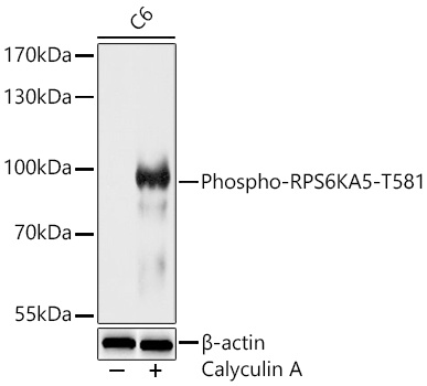
Western blot analysis of lysates from C6 cells using Phospho-RPS6KA5-T581 Rabbit pAb (CABP1197) at 1:1000 dilution. C6 cells were treated with Calyculin A (100 nM) at 37℃ for 30 minutes after serum-starvation overnight. Secondary antibody: HRP-conjugated Goat anti-Rabbit IgG (H+L) (CABS014) at 1:10000 dilution. Lysates/proteins: 25 μg per lane. Blocking buffer: 3% nonfat dry milk in TBST. Detection: ECL Basic Kit (AbGn00020). Exposure time: 60s.