The PI3 Kinase p110 delta Monoclonal Antibody (CAB19742) is a high-quality antibody developed for reliable detection and analysis of target proteins. This antibody, produced in rabbits, is highly specific to human samples and has been validated for use in Western blot applications. By binding to the p110 Delta protein, this antibody enables accurate detection and analysis in a variety of cell types, making it ideal for studies in cancer research and immunology.The PI3 Kinase pathway, particularly the p110 Delta isoform, plays a critical role in cell growth, proliferation, and survival.
This antibody is validated for use in WB, IHC-P, ELISA applications and has demonstrated reactivity against Human, Mouse, Rat samples.
Product Name:
PI3 Kinase p110 delta Monoclonal Antibody
SKU:
CAB19742
Size:
20μL, 100μL
Reactivity:
Human, Mouse, Rat
Clone Number:
ARC2268
Conjugate:
Unconjugated
Immunogen:
Synthetic peptide. This information is considered to be commercially sensitive.
Phosphoinositide 3-kinases (PI3Ks) phosphorylate inositol lipids and are involved in the immune response. The protein encoded by this gene is a class I PI3K found primarily in leukocytes. Like other class I PI3Ks (p110-alpha p110-beta, and p110-gamma), the encoded protein binds p85 adapter proteins and GTP-bound RAS. However, unlike the other class I PI3Ks, this protein phosphorylates itself, not p85 protein.
Purification Method
Affinity purification
Gene ID
5293
Buffer Information
Store at -20℃. Avoid freeze / thaw cycles. Buffer: PBS containing 50% glycerol and 0.05% BSA, preserved with proclin300 or sodium azide, pH 7.3.
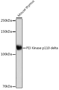
Western blot analysis of lysates from Mouse thymus, using PI3 Kinase p110 delta Rabbit mAb (CAB19742) at 1:1000 dilution incubated overnight at 4℃. Secondary antibody: HRP-conjugated Goat anti-Rabbit IgG (H+L) (CABS014) at 1:10000 dilution. Lysates/proteins: 25 μg per lane. Blocking buffer: 3% nonfat dry milk in TBST. Detection: ECL Basic Kit (AbGn00020). Exposure time: 180 s.
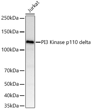
Western blot analysis of lysates from Jurkat cells using PI3 Kinase p110 delta Rabbit mAb (CAB19742) at 1:1000 dilution incubated at room temperature for 1.5 hours. Secondary antibody: HRP-conjugated Goat anti-Rabbit IgG (H+L) (CABS014) at 1:10000 dilution. Lysates/proteins: 25 μg per lane. Blocking buffer: 3% nonfat dry milk in TBST. Detection: ECL Basic Kit (AbGn00020). Exposure time: 20 s.
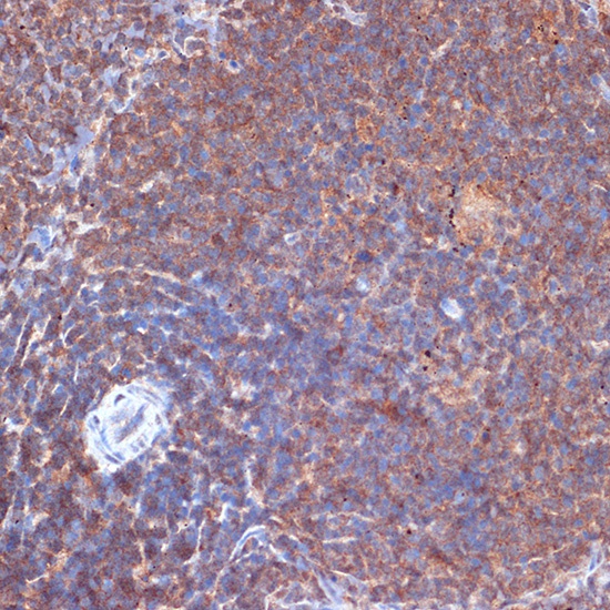
Immunohistochemistry analysis of paraffin-embedded Rat spleen using PI3 Kinase p110 delta Rabbit mAb (CAB19742) at dilution of 1:100 (40x lens). Microwave antigen retrieval performed with 0.01M Tris/EDTA Buffer (pH 9.0) prior to IHC staining.