The PIAS1+PIAS2 Monoclonal Antibody (CAB9670) is a high-quality antibody developed for reliable detection and analysis of target proteins. This gene encodes a member of the protein inhibitor of activated STAT (PIAS) family. PIAS proteins function as SUMO E3 ligases and play important roles in many cellular processes by mediating the sumoylation of target proteins. This protein plays a central role as a transcriptional coregulator of numerous cellular pathways includign the STAT1 and nuclear factor kappaB pathways. Alternate splicing results in multiple transcript variants. [provided by RefSeq, Mar 2016]
This antibody is validated for use in WB, IHC-P, ELISA applications and has demonstrated reactivity against Human, Mouse, Rat samples.
Product Name:
PIAS1+PIAS2 Monoclonal Antibody
SKU:
CAB9670
Size:
100μL, 20μL
Reactivity:
Human, Mouse, Rat
Clone Number:
ARC1690
Conjugate:
Unconjugated
Immunogen:
Synthetic peptide. This information is considered to be commercially sensitive.
Tested Applications:
WBIHC-PELISA
Recommended Dilution:
WB
1:500 - 1:2000
IHC-P
1:50 - 1:200
ELISA
Recommended starting concentration is 1 μg/mL. Please optimize the concentration based on your specific assay requirements.
Synonyms:
DDXBP1, GBP, GU/RH-II, ZMIZ3, PIAS1+PIAS2
Positive Sample:
HepG2, Mouse brain
Cellular Localization:
Nuclear Speck, Nucleoplasm, Nucleus, Pml Body.
Calculated MW:
76kDa
Observed MW:
76kDa
This gene encodes a member of the protein inhibitor of activated STAT (PIAS) family. PIAS proteins function as SUMO E3 ligases and play important roles in many cellular processes by mediating the sumoylation of target proteins. This protein plays a central role as a transcriptional coregulator of numerous cellular pathways includign the STAT1 and nuclear factor kappaB pathways. Alternate splicing results in multiple transcript variants. [provided by RefSeq, Mar 2016]
Purification Method
Affinity purification
Gene ID
8554 9063
RRID
AB_2863756
Buffer Information
Store at -20℃. Avoid freeze / thaw cycles. Buffer: PBS containing 50% glycerol and 0.05% BSA, preserved with proclin300 or sodium azide, pH 7.3.
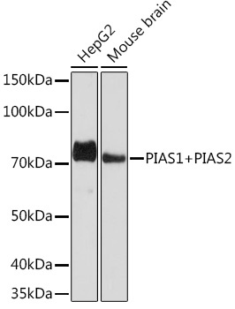
Western blot analysis of various lysates using PIAS1+PIAS2 Rabbit mAb (CAB9670) at 1:1000 dilution. Secondary antibody: HRP-conjugated Goat anti-Rabbit IgG (H+L) (AS014) at 1:10000 dilution. Lysates/proteins: 25μg per lane. Blocking buffer: 3% nonfat dry milk in TBST. Detection: ECL Basic Kit (AbGn00020). Exposure time: 90s.
Immunohistochemistry analysis of paraffin-embedded Human thyroid tissue using PIAS1+PIAS2 Rabbit mAb (CAB9670) at a dilution of 1:200 (40x lens). High pressure antigen retrieval was performed with 0.01 M Tris-EDTA buffer (pH 9.0) prior to IHC staining.
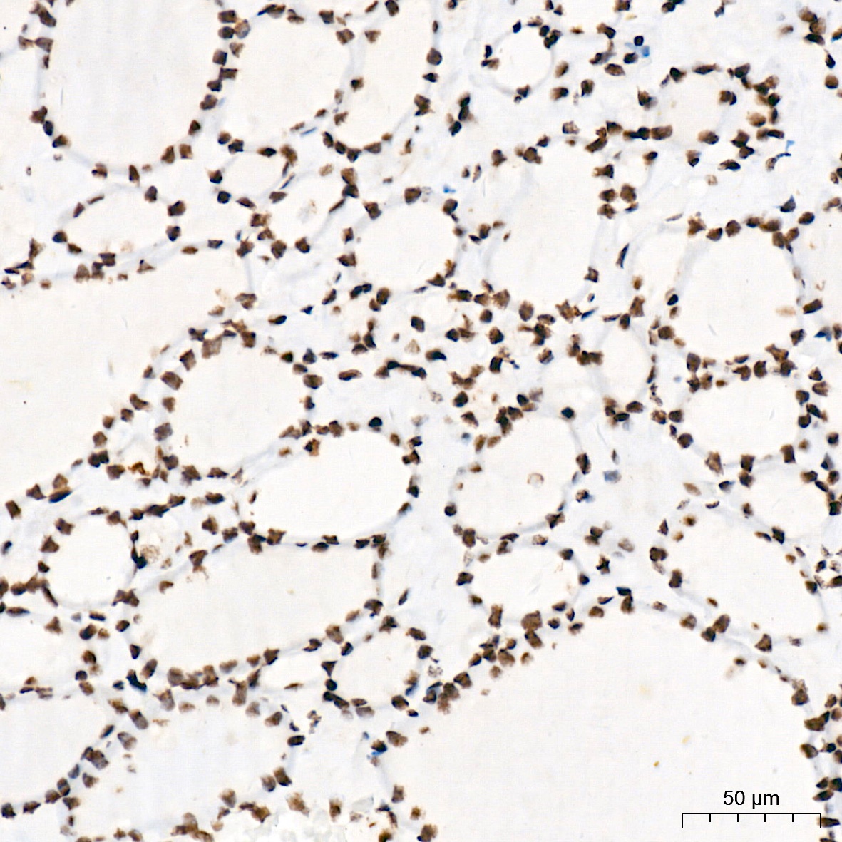
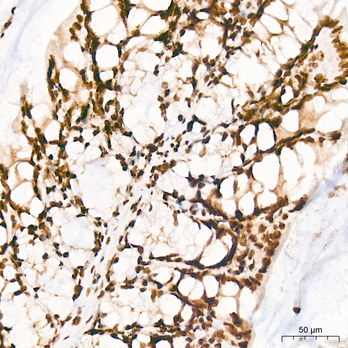
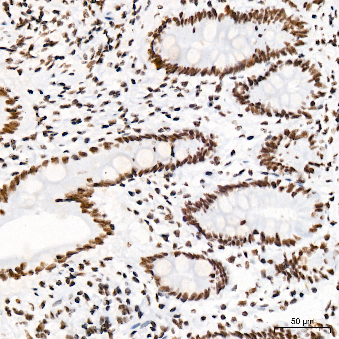
Immunohistochemistry analysis of paraffin-embedded Mouse colon tissue using PIAS1+PIAS2 Rabbit mAb (CAB9670) at a dilution of 1:200 (40x lens). High pressure antigen retrieval was performed with 0.01 M Tris-EDTA buffer (pH 9.0) prior to IHC staining.
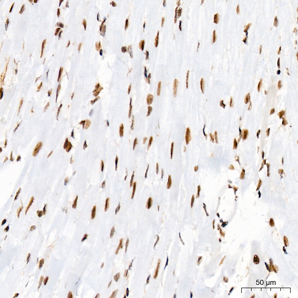
Immunohistochemistry analysis of paraffin-embedded Mouse heart tissue using PIAS1+PIAS2 Rabbit mAb (CAB9670) at a dilution of 1:200 (40x lens). High pressure antigen retrieval was performed with 0.01 M Tris-EDTA buffer (pH 9.0) prior to IHC staining.
Immunohistochemistry analysis of paraffin-embedded Human colon tissue using PIAS1+PIAS2 Rabbit mAb (CAB9670) at a dilution of 1:200 (40x lens). High pressure antigen retrieval was performed with 0.01 M Tris-EDTA buffer (pH 9.0) prior to IHC staining.
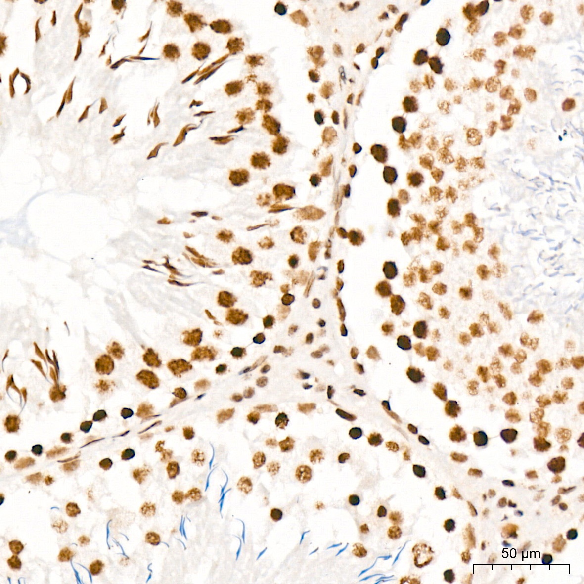
Immunohistochemistry analysis of paraffin-embedded Rat testis tissue using PIAS1+PIAS2 Rabbit mAb (CAB9670) at a dilution of 1:200 (40x lens). High pressure antigen retrieval was performed with 0.01 M Tris-EDTA buffer (pH 9.0) prior to IHC staining.