The PLOD3 Monoclonal Antibody (CAB21066) is a high-quality antibody developed for reliable detection and analysis of target proteins. This antibody, developed in rabbits, demonstrates high reactivity with human samples and has been validated for use in Western blot applications. By binding specifically to the PLod3 protein, this antibody enables precise detection and analysis in various cell types, making it an essential component for investigations in the fields of biochemistry and molecular biology.PLod3, also known as procollagen-lysine, 2-oxoglutarate 5-dioxygenase 3, plays a crucial role in collagen biosynthesis by catalyzing the hydroxylation of lysine residues.
This antibody is validated for use in WB, ELISA applications and has demonstrated reactivity against Human, Mouse samples.
Product Name:
PLOD3 Monoclonal Antibody
SKU:
CAB21066
Size:
20μL, 100μL
Reactivity:
Human, Mouse
Clone Number:
ARC52014
Conjugate:
Unconjugated
Immunogen:
Recombinant protein (or fragment).This information is considered to be commercially sensitive.
The protein encoded by this gene is a membrane-bound homodimeric enzyme that is localized to the cisternae of the rough endoplasmic reticulum. The enzyme (cofactors iron and ascorbate) catalyzes the hydroxylation of lysyl residues in collagen-like peptides. The resultant hydroxylysyl groups are attachment sites for carbohydrates in collagen and thus are critical for the stability of intermolecular crosslinks. Some patients with Ehlers-Danlos syndrome type VIB have deficiencies in lysyl hydroxylase activity.
Purification Method
Affinity purification
Gene ID
8985
Buffer Information
Store at -20℃. Avoid freeze / thaw cycles. Buffer: PBS containing 50% glycerol and 0.05% BSA, preserved with proclin300 or sodium azide, pH 7.3.
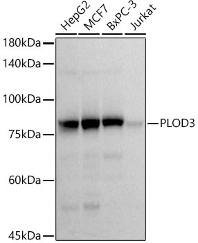
Western blot analysis of various lysates using PLOD3 Rabbit mAb (CAB21066) at1:1000 dilution. Secondary antibody: HRP-conjugated Goat anti-Rabbit IgG (H+L) (CABS014) at1:10000 dilution. Lysates/proteins: 25μg per lane. Blocking buffer: 3% nonfat dry milk in TBST. Detection: ECL Basic Kit (AbGn00020). Exposure time: 3s.
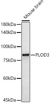
Western blot analysis of lysates from Mouse brain, using PLOD3 Rabbit mAb (CAB21066) at1:1000 dilution. Secondary antibody: HRP-conjugated Goat anti-Rabbit IgG (H+L) (CABS014) at1:10000 dilution. Lysates/proteins: 25μg per lane. Blocking buffer: 3% nonfat dry milk in TBST. Detection: ECL Basic Kit (AbGn00020). Exposure time: 180s.
 at 1:10000 dilution. Lysates/proteins: 25ug per lane. Blocking buffer: 3% nonfat dry milk in TBST. Detection: ECL Basic Kit. Exposure time: 180s.")
. Perform high pressure antigen retrieval with 10 mM citrate buffer pH 6. 0 before commencing with IHC staining protocol.")
 at 1:10000 dilution. Lysates/proteins: 25ug per lane. Blocking buffer: 3% nonfat dry milk in TBST. Detection: ECL Basic Kit. Exposure time: 3s.")
 at 1:10000 dilution. Lysates/proteins: 25ug per lane. Blocking buffer: 3% nonfat dry milk in TBST. Detection: ECL Basic Kit. Exposure time: 180s.")


