The Proteinase K Antibody (CAB20453) is a high-quality antibody developed for reliable detection and analysis of target proteins. Proteinase K is an endopeptidase belonging to the subtilisin group of serine proteases. Proteinase K was isolated from the mold (Parengyodontium album or previously known as Tritirachium album). Proteinase K has five cysteines. Four of the cysteine residues form two disulfide bonds (34-124 and 179-248, respectively) and one lies below one of the catalytic triads. Proteinase K often binds two calcium ions required for full enzymatic activity. In molecular biology laboratories, proteinase K is useful during preparation of DNA or RNA samples by degrading and inactivating proteins.
This antibody is validated for use in WB, ELISA applications and has demonstrated reactivity against Fungi samples.
Product Name:
Proteinase K Antibody
SKU:
CAB20453
Size:
100μL, 20μL
Reactivity:
Fungi
Conjugate:
Unconjugated
Immunogen:
Recombinant protein (or fragment).This information is considered to be commercially sensitive.
Tested Applications:
WBELISA
Recommended Dilution:
WB
1:500 - 1:1000
ELISA
Recommended starting concentration is 1 μg/mL. Please optimize the concentration based on your specific assay requirements.
Positive Sample:
Proteinase K
Observed MW:
60kDa
Proteinase K is an endopeptidase belonging to the subtilisin group of serine proteases. Proteinase K was isolated from the mold (Parengyodontium album or previously known as Tritirachium album). Proteinase K has five cysteines. Four of the cysteine residues form two disulfide bonds (34-124 and 179-248, respectively) and one lies below one of the catalytic triads. Proteinase K often binds two calcium ions required for full enzymatic activity. In molecular biology laboratories, proteinase K is useful during preparation of DNA or RNA samples by degrading and inactivating proteins.
Purification Method
Affinity purification
Buffer Information
Store at -20℃. Avoid freeze / thaw cycles. Buffer: PBS containing 50% glycerol, preserved with proclin300 or sodium azide, pH 7.3.
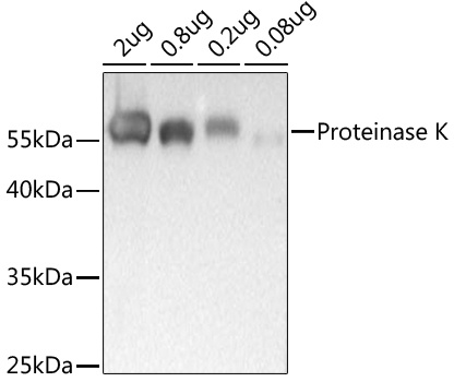
Western blot analysis of recombinant Proteinase K Protein using Proteinase K Rabbit pAb (CAB20453) at 1:1000 dilution incubated overnight at 4℃. Secondary antibody: HRP-conjugated Goat anti-Rabbit IgG (H+L) (AS014) at 1:10000 dilution. Lysates/proteins: 25μg per lane. Blocking buffer: 3% nonfat dry milk in TBST. Detection: ECL Basic Kit (AbGn00020). Exposure time: .