The SNRPA1 Monoclonal Antibody (CAB0647) is a high-quality antibody developed for reliable detection and analysis of target proteins. SNRPA1, also known as U1 small nuclear ribonucleoprotein A, is a key component of the spliceosome complex involved in pre-mRNA splicing. This antibody is raised in rabbits and is highly reactive with human samples. It has been validated for use in Western blot applications, allowing for the detection and analysis of SNRPA1 in various cell types. This makes it an ideal tool for studies in molecular biology and RNA splicing mechanisms.
This antibody is validated for use in WB, IHC-P, IP, ELISA applications and has demonstrated reactivity against Human, Mouse, Rat samples.
Product Name:
SNRPA1 Monoclonal Antibody
SKU:
CAB0647
Size:
20μL, 100μL
Reactivity:
Human, Mouse, Rat
Clone Number:
ARC2532
Conjugate:
Unconjugated
Immunogen:
Synthetic peptide. This information is considered to be commercially sensitive.
Enables RNA binding activity. Involved in mRNA splicing, via spliceosome and spermatogenesis. Located in nuclear speck. Part of U2-type catalytic step 2 spliceosome and U2-type precatalytic spliceosome. Implicated in connective tissue disease.
Purification Method
Affinity purification
Gene ID
6627
Buffer Information
Store at -20℃. Avoid freeze / thaw cycles. Buffer: PBS containing 50% glycerol and 0.05% BSA, preserved with proclin300 or sodium azide, pH 7.3.
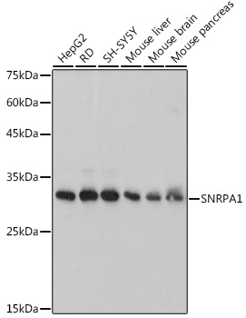
Western blot analysis of various lysates, using SNRPA1 Rabbit mAb (CAB0647) at 1:1000 dilution. Secondary antibody: HRP-conjugated Goat anti-Rabbit IgG (H+L) (CABS014) at 1:10000 dilution. Lysates/proteins: 25μg per lane. Blocking buffer: 3% nonfat dry milk in TBST. Detection: ECL Basic Kit (AbGn00020). Exposure time: 3s.
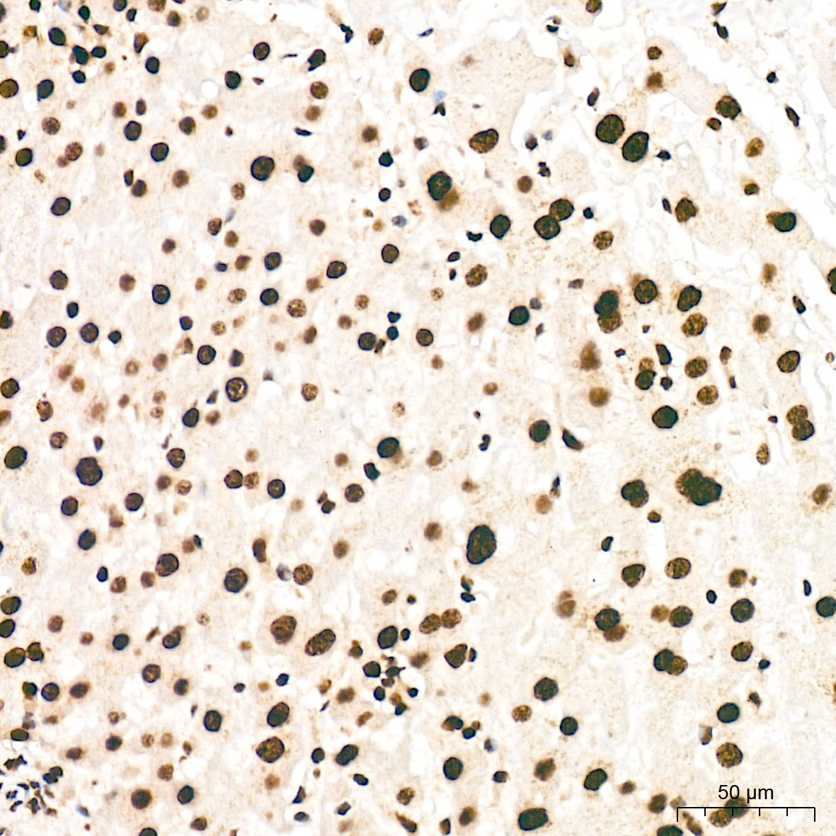
Immunohistochemistry analysis of paraffin-embedded Human liver tissue using SNRPA1 Rabbit mAb (CAB0647) at a dilution of 1:200 (40x lens). High pressure antigen retrieval was performed with 0.01 M citrate buffer (pH 6.0) prior to IHC staining.
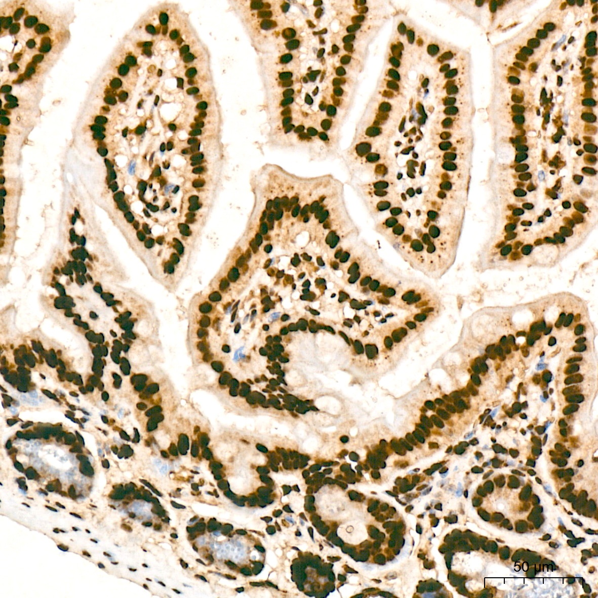
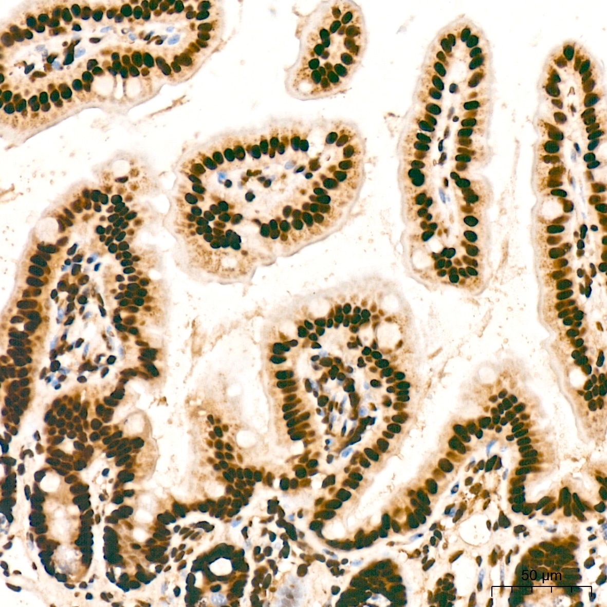
Immunohistochemistry analysis of paraffin-embedded Mouse intestin tissue using SNRPA1 Rabbit mAb (CAB0647) at a dilution of 1:200 (40x lens). High pressure antigen retrieval was performed with 0.01 M citrate buffer (pH 6.0) prior to IHC staining.
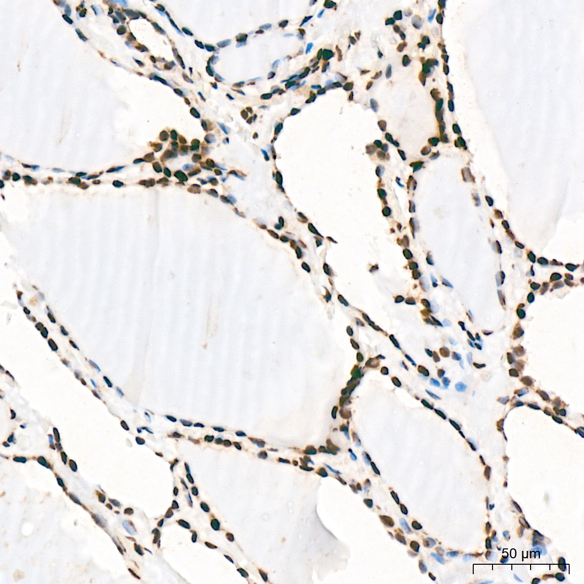
Immunohistochemistry analysis of paraffin-embedded Rat intestine tissue using SNRPA1 Rabbit mAb (CAB0647) at a dilution of 1:200 (40x lens). High pressure antigen retrieval was performed with 0.01 M citrate buffer (pH 6.0) prior to IHC staining.
Immunohistochemistry analysis of paraffin-embedded Rat intestine tissue using SNRPA1 Rabbit mAb (CAB0647) at a dilution of 1:200 (40x lens). High pressure antigen retrieval was performed with 0.01 M citrate buffer (pH 6.0) prior to IHC staining.
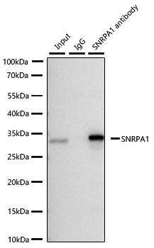
Immunoprecipitation of SNRPA1 from 300 µg extracts of Hep G2 cells was performed using 3 µg of SNRPA1 Rabbit mAb (CAB0647). Rabbit Control IgG (AC005) was used to precipitate the Control IgG sample. IP samples were eluted with 1× Laemmli Buffer. The Input lane represents 10% of the total input. Western blot analysis of immunoprecipitates was conducted using SNRPA1 Rabbit mAb (CAB0647) at a dilution of 1:5000.