The STK33 Monoclonal Antibody (CAB19803) is a high-quality antibody developed for reliable detection and analysis of target proteins. This antibody, generated in mice, is highly specific and reacts with human samples, making it ideal for use in Western blot and immunofluorescence applications. By targeting the STK33 protein, this antibody allows for the detection and analysis of STK33 expression in various cell types, offering insights into its role in cellular processes.
This antibody is validated for use in WB, IF/ICC, ELISA applications and has demonstrated reactivity against Human, Mouse samples.
Product Name:
STK33 Monoclonal Antibody
SKU:
CAB19803
Size:
20μL, 100μL
Reactivity:
Human, Mouse
Clone Number:
ARC2330
Conjugate:
Unconjugated
Immunogen:
Recombinant protein (or fragment).This information is considered to be commercially sensitive.
Recommended starting concentration is 1 μg/mL. Please optimize the concentration based on your specific assay requirements.
Synonyms:
STK33
Positive Sample:
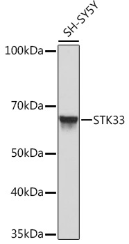
SH-SY5Y
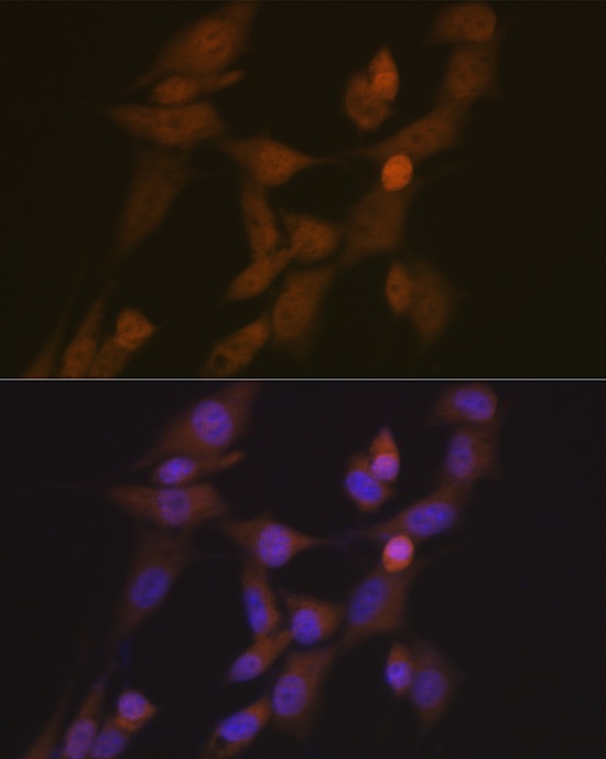
Cellular Localization:
Cytoplasm, Nucleus, Perinuclear Region Of Cytoplasm.
Calculated MW:
58kDa
Observed MW:
58kDa
Serine/threonine protein kinase (STK33) is a 524-residue kinase that belongs to the calcium/calmodulin-dependent family of kinases. STK33 exhibits differential expression in normal and malignant cancer tissue, including hepatocellular carcinoma. STK33 expression is required in the context of mutant KRAS, the most commonly mutated human oncogene, to maintain for cellular viability and proliferation. STK33 depletion via modulation of interacting proteins results in proteasome-mediated degradation of STK33 in human cancer cells, triggering apoptosis.
Purification Method
Affinity purification
Gene ID
65975
Buffer Information
Store at -20℃. Avoid freeze / thaw cycles. Buffer: PBS containing 50% glycerol and 0.05% BSA, preserved with proclin300 or sodium azide, pH 7.3.
Western blot analysis of lysates from SH-SY5Y cells, using STK33 Rabbit mAb (CAB19803) at 1:1000 dilution. Secondary antibody: HRP-conjugated Goat anti-Rabbit IgG (H+L) (CABS014) at 1:10000 dilution. Lysates/proteins: 25μg per lane. Blocking buffer: 3% nonfat dry milk in TBST. Detection: ECL Basic Kit (AbGn00020). Exposure time: 10s.
Immunofluorescence analysis of NIH/3T3 cells using STK33 Rabbit mAb (CAB19803) at dilution of 1:100 (40x lens). Secondary antibody: Cy3-conjugated Goat anti-Rabbit IgG (H+L) (CABS007) at 1:500 dilution. Blue: DAPI for nuclear staining.