The SUOX Monoclonal Antibody (CAB0733) is a high-quality antibody developed for reliable detection and analysis of target proteins. The antibody, produced in rabbits, demonstrates high reactivity with human samples and is validated for use in Western blot applications. By binding specifically to the SUOX protein, this antibody allows for the detection and analysis of SUOX in a variety of cell types, making it an excellent choice for studies in biochemistry and metabolic disorders.SUOX plays a crucial role in the detoxification of sulphur-containing compounds in the body, making it a key player in maintaining proper sulphur balance and preventing toxicity.
This antibody is validated for use in WB, ELISA applications and has demonstrated reactivity against Rat samples.
Product Name:
SUOX Monoclonal Antibody
SKU:
CAB0733
Size:
20μL, 100μL
Reactivity:
Rat
Clone Number:
ARC2535
Conjugate:
Unconjugated
Immunogen:
Synthetic peptide. This information is considered to be commercially sensitive.
Recommended starting concentration is 1 μg/mL. Please optimize the concentration based on your specific assay requirements.
Synonyms:
SUOX, sulfite oxidase
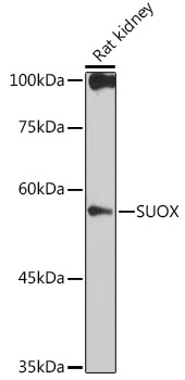
Positive Sample:
Rat kidney
Cellular Localization:
Mitochondrion Intermembrane Space.
Calculated MW:
60kDa
Observed MW:
60kDa
Sulfite oxidase is a homodimeric protein localized to the intermembrane space of mitochondria. Each subunit contains a heme domain and a molybdopterin-binding domain. The enzyme catalyzes the oxidation of sulfite to sulfate, the final reaction in the oxidative degradation of the sulfur amino acids cysteine and methionine. Sulfite oxidase deficiency results in neurological abnormalities which are often fatal at an early age. Alternative splicing results in multiple transcript variants encoding identical proteins.
Purification Method
Affinity purification
Gene ID
6821
Buffer Information
Store at -20℃. Avoid freeze / thaw cycles. Buffer: PBS containing 50% glycerol and 0.05% BSA, preserved with proclin300 or sodium azide, pH 7.3.
Western blot analysis of lysates from Rat kidney, using SUOX Rabbit mAb (CAB0733) at 1:1000 dilution. Secondary antibody: HRP-conjugated Goat anti-Rabbit IgG (H+L) (CABS014) at 1:10000 dilution. Lysates/proteins: 25μg per lane. Blocking buffer: 3% nonfat dry milk in TBST. Detection: ECL Basic Kit (AbGn00020). Exposure time: 30s.