The TAB1 Monoclonal Antibody (CAB3306) is a high-quality antibody developed for reliable detection and analysis of target proteins. This antibody, produced in rabbits, has high reactivity with human samples and has been validated for use in Western blot applications. By binding to the TAB1 protein, this antibody allows for the detection and analysis of TAB1 in various cell types, making it an excellent choice for studies in molecular biology and signal transduction research.
This antibody is validated for use in WB, IF/ICC, ELISA applications and has demonstrated reactivity against Human, Mouse, Rat samples.
Product Name:
TAB1 Monoclonal Antibody
SKU:
CAB3306
Size:
20μL, 100μL
Reactivity:
Human, Mouse, Rat
Clone Number:
ARC1943
Conjugate:
Unconjugated
Immunogen:
Synthetic peptide. This information is considered to be commercially sensitive.
Recommended starting concentration is 1 μg/mL. Please optimize the concentration based on your specific assay requirements.
Synonyms:
3'-Tab1, MAP3K7IP1, TAB1
Positive Sample:
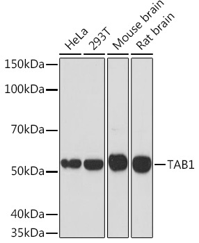
HeLa, 293T, Mouse brain, Rat brain
Cellular Localization:
Cytosol, Nuclear Speck, Nucleus.
Calculated MW:
55kDa
Observed MW:
55kDa
The protein encoded by this gene was identified as a regulator of the MAP kinase kinase kinase MAP3K7/TAK1, which is known to mediate various intracellular signaling pathways, such as those induced by TGF beta, interleukin 1, and WNT-1. This protein interacts and thus activates TAK1 kinase. It has been shown that the C-terminal portion of this protein is sufficient for binding and activation of TAK1, while a portion of the N-terminus acts as a dominant-negative inhibitor of TGF beta, suggesting that this protein may function as a mediator between TGF beta receptors and TAK1. This protein can also interact with and activate the mitogen-activated protein kinase 14 (MAPK14/p38alpha), and thus represents an alternative activation pathway, in addition to the MAPKK pathways, which contributes to the biological responses of MAPK14 to various stimuli. Alternatively spliced transcript variants encoding distinct isoforms have been reported.
Purification Method
Affinity purification
Gene ID
10454
RRID
AB_2863038
Buffer Information
Store at -20℃. Avoid freeze / thaw cycles. Buffer: PBS containing 50% glycerol and 0.05% BSA, preserved with proclin300 or sodium azide, pH 7.3.
Western blot analysis of various lysates using TAB1 Rabbit mAb (CAB3306) at 1:1000 dilution. Secondary antibody: HRP-conjugated Goat anti-Rabbit IgG (H+L) (CABS014) at 1:10000 dilution. Lysates/proteins: 25μg per lane. Blocking buffer: 3% nonfat dry milk in TBST. Detection: ECL Basic Kit (AbGn00020). Exposure time: 90s.
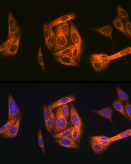
Immunofluorescence analysis of U-2 OS cells using TAB1 Rabbit mAb (CAB3306) at dilution of 1:100 (40x lens). Secondary antibody: Cy3-conjugated Goat anti-Rabbit IgG (H+L) (CABS007) at 1:500 dilution. Blue: DAPI for nuclear staining.