The TAB3 Monoclonal Antibody (CAB19824) is a high-quality antibody developed for reliable detection and analysis of target proteins. This antibody, produced in rabbits, exhibits high reactivity with human samples and has been validated for use in Western blot applications. By specifically binding to TAB3, this antibody enables the accurate detection and analysis of the protein in various cell types, making it an invaluable asset for immunology and cancer research projects.TAB3, also known as TGF-beta-activated kinase-binding protein 3, is a crucial mediator of pro-inflammatory signaling cascades, making it a promising target for therapeutic intervention in diseases characterized by dysregulated immune responses.
This antibody is validated for use in WB, ELISA applications and has demonstrated reactivity against Human, Mouse samples.
Product Name:
TAB3 Monoclonal Antibody
SKU:
CAB19824
Size:
20μL, 100μL
Reactivity:
Human, Mouse
Clone Number:
ARC2350
Conjugate:
Unconjugated
Immunogen:
Synthetic peptide. This information is considered to be commercially sensitive.
Recommended starting concentration is 1 μg/mL. Please optimize the concentration based on your specific assay requirements.
Synonyms:
NAP1, MAP3K7IP3, TAB3
Positive Sample:
HeLa, 293T, HepG2, Mouse liver
Cellular Localization:
Cytosol, Extracellular Exosome, Plasma Membrane.
Calculated MW:
79kDa
Observed MW:
100kDa
The product of this gene functions in the NF-kappaB signal transduction pathway. The encoded protein, and the similar and functionally redundant protein MAP3K7IP2/TAB2, forms a ternary complex with the protein kinase MAP3K7/TAK1 and either TRAF2 or TRAF6 in response to stimulation with the pro-inflammatory cytokines TNF or IL-1. Subsequent MAP3K7/TAK1 kinase activity triggers a signaling cascade leading to activation of the NF-kappaB transcription factor. The human genome contains a related pseudogene. Alternatively spliced transcript variants have been described, but their biological validity has not been determined.
Purification Method
Affinity purification
Gene ID
257397
Buffer Information
Store at -20℃. Avoid freeze / thaw cycles. Buffer: PBS containing 50% glycerol and 0.05% BSA, preserved with proclin300 or sodium azide, pH 7.3.
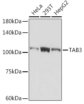
Western blot analysis of various lysates using TAB3 Rabbit mAb (CAB19824) at 1:1000 dilution. Secondary antibody: HRP-conjugated Goat anti-Rabbit IgG (H+L) (CABS014) at 1:10000 dilution. Lysates/proteins: 25μg per lane. Blocking buffer: 3% nonfat dry milk in TBST. Detection: ECL Basic Kit (AbGn00020). Exposure time: 5s.
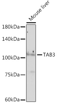
Western blot analysis of lysates from mouse liver using TAB3 Rabbit mAb (CAB19824) at 1:1000 dilution. Secondary antibody: HRP-conjugated Goat anti-Rabbit IgG (H+L) (CABS014) at 1:10000 dilution. Lysates/proteins: 25 μg per lane. Blocking buffer: 3% nonfat dry milk in TBST. Detection: ECL Enhanced Kit (AbGn00021). Exposure time: 180s.