The TARBP2 Monoclonal Antibody (CAB9105) is a high-quality antibody developed for reliable detection and analysis of target proteins. HIV-1, the causative agent of acquired immunodeficiency syndrome (AIDS), contains an RNA genome that produces a chromosomally integrated DNA during the replicative cycle. Activation of HIV-1 gene expression by the transactivator Tat is dependent on an RNA regulatory element (TAR) located downstream of the transcription initiation site. The protein encoded by this gene binds between the bulge and the loop of the HIV-1 TAR RNA regulatory element and activates HIV-1 gene expression in synergy with the viral Tat protein. Alternative splicing results in multiple transcript variants encoding different isoforms. This gene also has a pseudogene. RRID AB_2863657 Gene ID 6895 Swiss Prot Synonym LOQS; TRBP; TRBP1; TRBP2; TARBP2
This antibody is validated for use in WB, IHC-P, IF/ICC, ELISA, IF-P applications and has demonstrated reactivity against Human, Mouse, Rat samples.
Product Name:
TARBP2 Monoclonal Antibody
SKU:
CAB9105
Size:
100μL, 20μL
Reactivity:
Human, Mouse, Rat
Clone Number:
ARC1792
Conjugate:
Unconjugated
Immunogen:
Synthetic peptide. This information is considered to be commercially sensitive.
Tested Applications:
WBIHC-PIF/ICCELISAIF-P
Recommended Dilution:
WB
1:500 - 1:2000
IF
/
ICC
1:50 - 1:200
IF-P
1:50 - 1:200
IHC-P
1:50 - 1:200
ELISA
Recommended starting concentration is 1 μg/mL. Please optimize the concentration based on your specific assay requirements.
Synonyms:
LOQS, TRBP, TRBP1, TRBP2, TARBP2
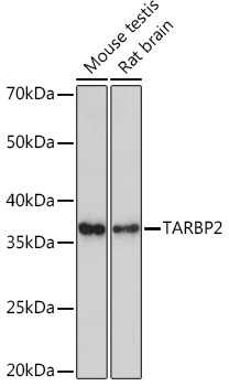
Positive Sample:
Mouse testis, Rat brain
Cellular Localization:
Cytoplasm, Nucleus, Perinuclear Region.
Calculated MW:
39kDa
Observed MW:
-
HIV-1, the causative agent of acquired immunodeficiency syndrome (AIDS), contains an RNA genome that produces a chromosomally integrated DNA during the replicative cycle. Activation of HIV-1 gene expression by the transactivator Tat is dependent on an RNA regulatory element (TAR) located downstream of the transcription initiation site. The protein encoded by this gene binds between the bulge and the loop of the HIV-1 TAR RNA regulatory element and activates HIV-1 gene expression in synergy with the viral Tat protein. Alternative splicing results in multiple transcript variants encoding different isoforms. This gene also has a pseudogene. RRID AB_2863657 Gene ID 6895 Swiss Prot Synonym LOQS; TRBP; TRBP1; TRBP2; TARBP2
Purification Method:
Affinity purification
Gene ID:
6895
RRID:
AB_2863657
Buffer Information:
Store at -20℃. Avoid freeze / thaw cycles. Buffer: PBS containing 50% glycerol and 0.05% BSA, preserved with proclin300 or sodium azide, pH 7.3.
Western blot analysis of various lysates using TARBP2 Rabbit mAb (CAB9105) at 1:1000 dilution. Secondary antibody: HRP-conjugated Goat anti-Rabbit IgG (H+L) (AS014) at 1:10000 dilution. Lysates/proteins: 25μg per lane. Blocking buffer: 3% nonfat dry milk in TBST. Detection: ECL Enhanced Kit (AbGn00021). Exposure time: 3min.
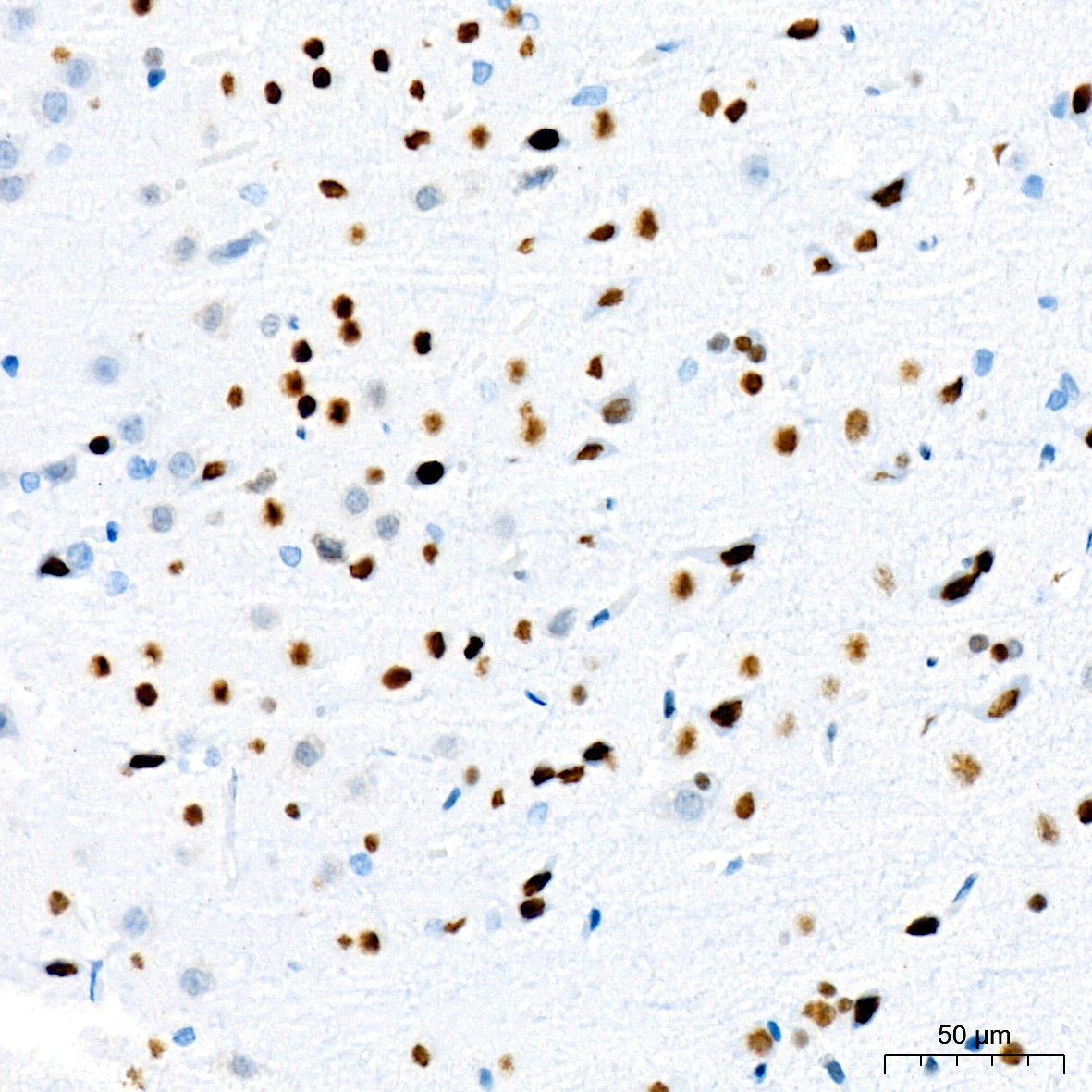
Immunohistochemistry analysis of paraffin-embedded Rat brain tissue using TARBP2 Rabbit mAb (CAB9105) at a dilution of 1:200 (40x lens). High pressure antigen retrieval was performed with 0.01 M citrate buffer (pH 6.0) prior to IHC staining.
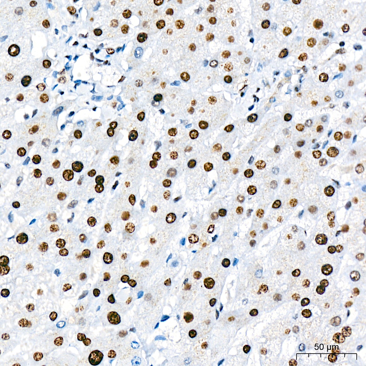
Immunohistochemistry analysis of paraffin-embedded Human liver tissue using TARBP2 Rabbit mAb (CAB9105) at a dilution of 1:200 (40x lens). High pressure antigen retrieval was performed with 0.01 M citrate buffer (pH 6.0) prior to IHC staining.
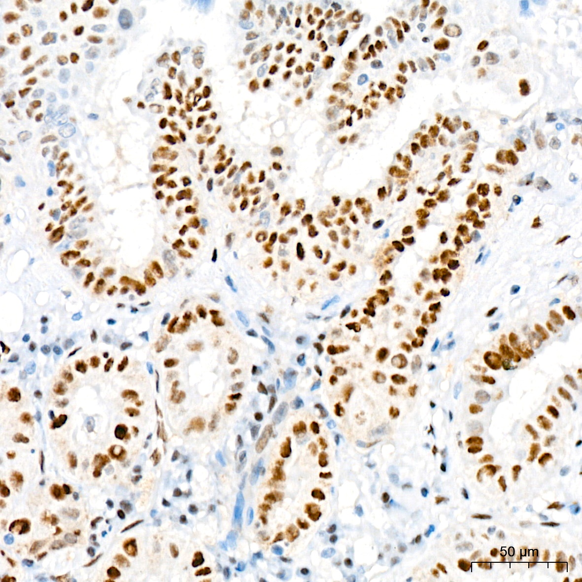
Immunohistochemistry analysis of paraffin-embedded Human thyroid cancer tissue using TARBP2 Rabbit mAb (CAB9105) at a dilution of 1:200 (40x lens). High pressure antigen retrieval was performed with 0.01 M citrate buffer (pH 6.0) prior to IHC staining.
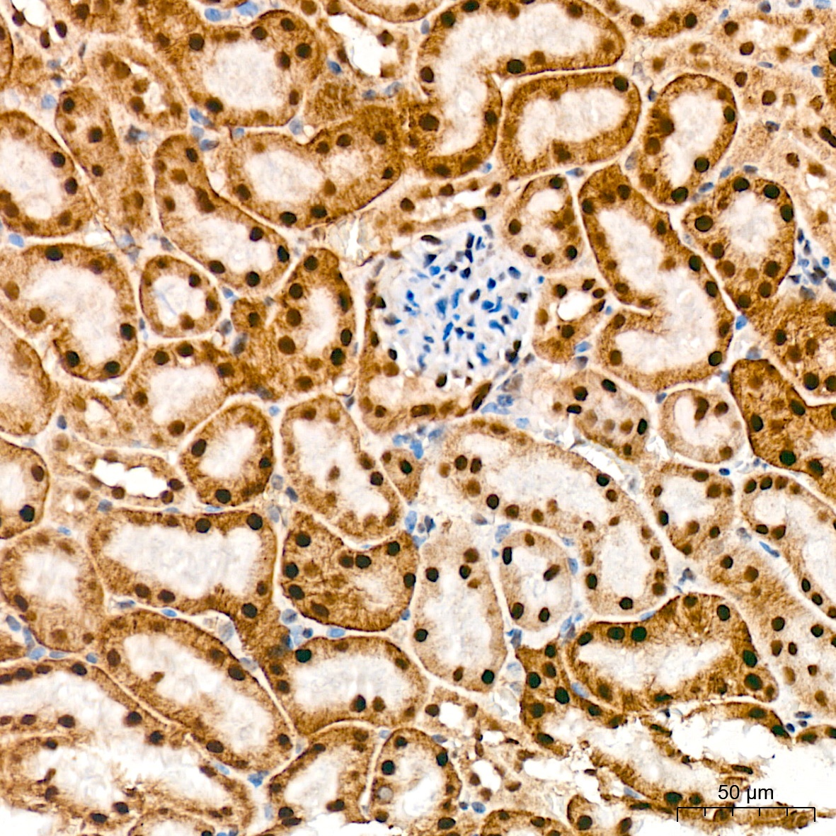
Immunohistochemistry analysis of paraffin-embedded Mouse kidney tissue using TARBP2 Rabbit mAb (CAB9105) at a dilution of 1:200 (40x lens). High pressure antigen retrieval was performed with 0.01 M citrate buffer (pH 6.0) prior to IHC staining.
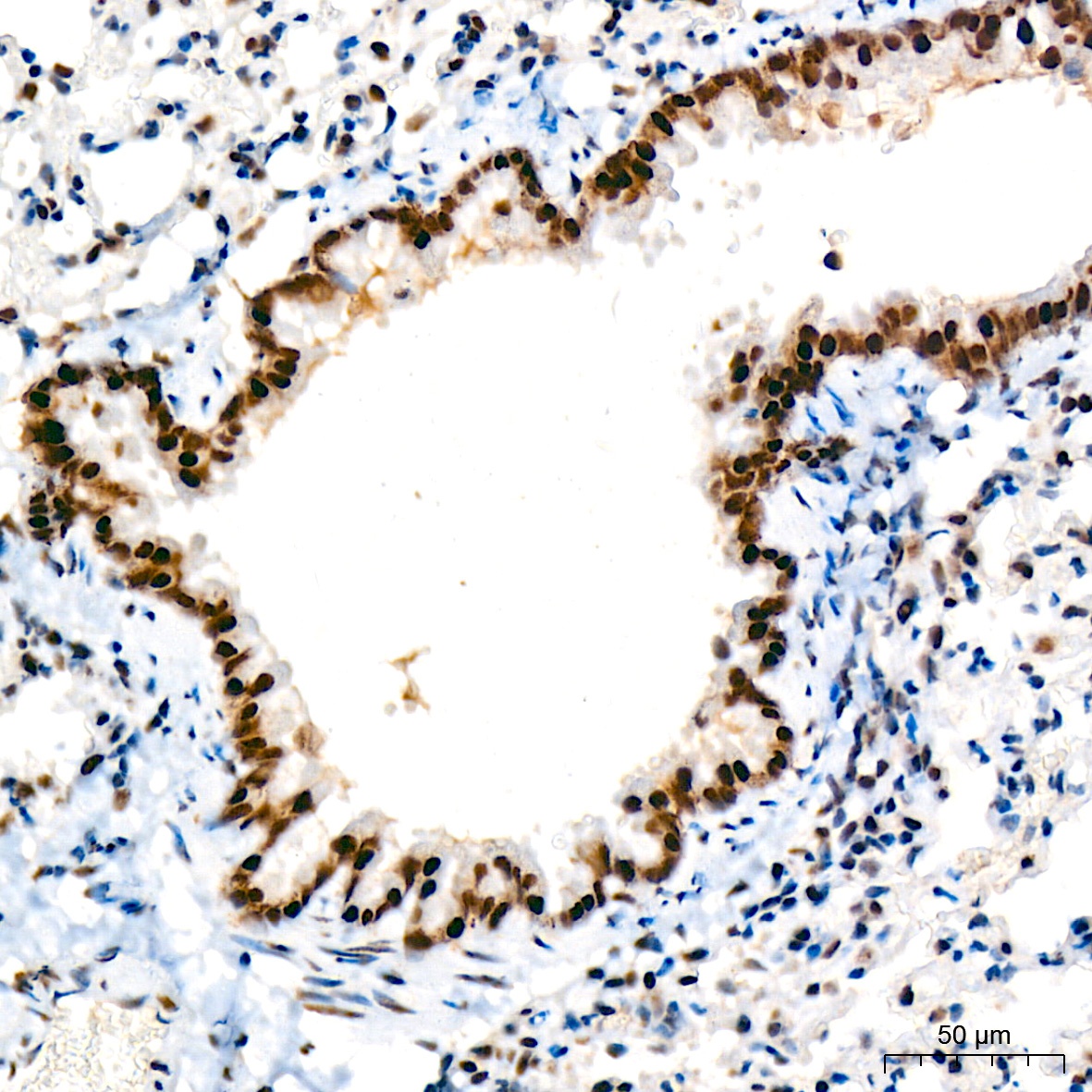
Immunohistochemistry analysis of paraffin-embedded Mouse lung tissue using TARBP2 Rabbit mAb (CAB9105) at a dilution of 1:200 (40x lens). High pressure antigen retrieval was performed with 0.01 M citrate buffer (pH 6.0) prior to IHC staining.
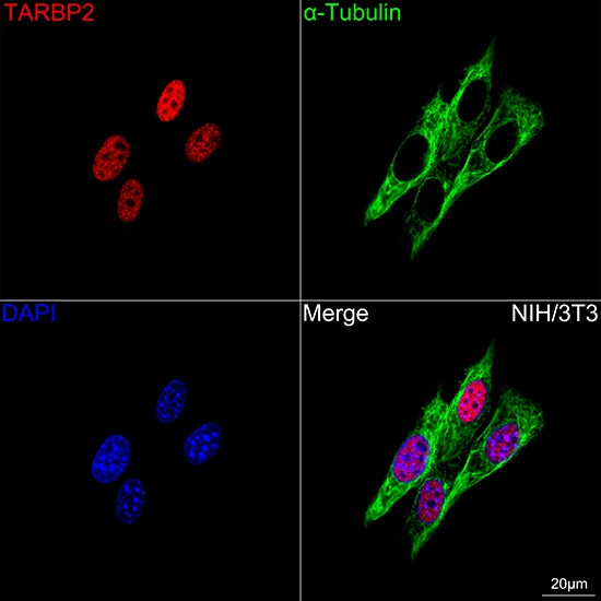
Confocal imaging of NIH/3T3 cells using TARBP2 Rabbit mAb (CAB9105, dilution 1:200) followed by a further incubation with Cy3 Goat Anti-Rabbit IgG (H+L) (AS007, dilution 1:500) (Red). The cells were counterstained with α-Tubulin Mouse mAb (AC012, dilution 1:400) followed by incubation with ABflo® 488-conjugated Goat Anti-Mouse IgG (H+L) Ab (AS076, dilution 1:500) (Green). DAPI was used for nuclear staining (Blue). Objective: 100x.
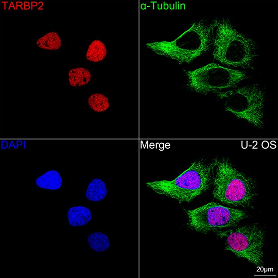
Confocal imaging of U-2 OS cells using TARBP2 Rabbit mAb (CAB9105,dilution 1:200) followed by a further incubation with Cy3 Goat Anti-Rabbit IgG (H+L) (AS007, dilution 1:500) (Red). The cells were counterstained with α-Tubulin Mouse mAb (AC012, dilution 1:400) followed by incubation with ABflo® 488-conjugated Goat Anti-Mouse IgG (H+L) Ab (AS076, dilution 1:500) (Green). DAPI was used for nuclear staining (Blue). Objective: 100x.