The TIMM50 Monoclonal Antibody (CAB1536) is a high-quality antibody developed for reliable detection and analysis of target proteins. This antibody, generated in rabbits, demonstrates strong reactivity with human samples and has been validated for use in Western blot applications. By binding specifically to the TIMM50 protein, researchers can accurately detect and analyze its expression in various cell types, making it an excellent tool for studies in mitochondrial biology and disease research.
This antibody is validated for use in WB, IHC-P, IF/ICC, ELISA, IF-P applications and has demonstrated reactivity against Human, Mouse, Rat samples.
Product Name:
TIMM50 Monoclonal Antibody
SKU:
CAB1536
Size:
20μL, 100μL
Reactivity:
Human, Mouse, Rat
Clone Number:
ARC1883
Conjugate:
Unconjugated
Immunogen:
Synthetic peptide. This information is considered to be commercially sensitive.
This gene encodes a subunit of the TIM23 inner mitochondrial membrane translocase complex. The encoded protein functions as the receptor subunit that recognizes the mitochondrial targeting signal, or presequence, on protein cargo that is destined for the mitochondrial inner membrane and matrix. This protein may also play a role in maintaining the membrane permeability barrier, and knockdown of this gene in human cells results in the release of cytochrome c and apoptosis.
Purification Method
Affinity purification
Gene ID
92609
RRID
AB_2861715
Buffer Information
Store at -20℃. Avoid freeze / thaw cycles. Buffer: PBS containing 50% glycerol and 0.05% BSA, preserved with proclin300 or sodium azide, pH 7.3.
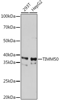
Western blot analysis of various lysates using TIMM50 Rabbit mAb (CAB1536) at 1:1000 dilution. Secondary antibody: HRP-conjugated Goat anti-Rabbit IgG (H+L) (CABS014) at 1:10000 dilution. Lysates/proteins: 25μg per lane. Blocking buffer: 3% nonfat dry milk in TBST. Detection: ECL Basic Kit (AbGn00020). Exposure time: 30s.
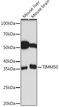
Western blot analysis of various lysates using TIMM50 Rabbit mAb (CAB1536) at 1:1000 dilution. Secondary antibody: HRP-conjugated Goat anti-Rabbit IgG (H+L) (CABS014) at 1:10000 dilution. Lysates/proteins: 25μg per lane. Blocking buffer: 3% nonfat dry milk in TBST. Detection: ECL Basic Kit (AbGn00020). Exposure time: 60s.
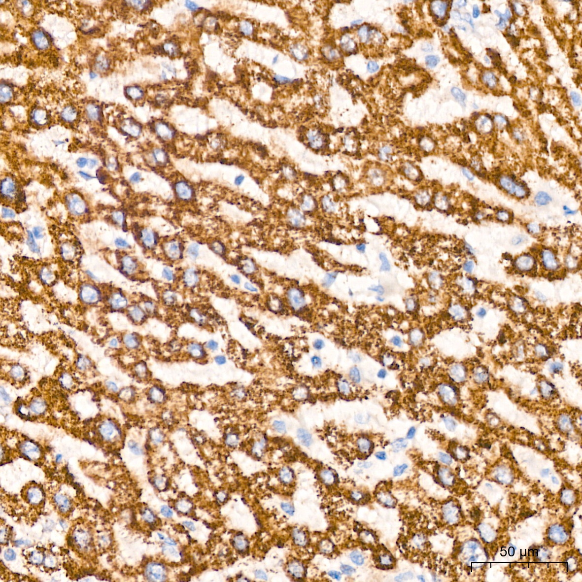
Immunohistochemistry analysis of paraffin-embedded Human liver tissue using TIMM50 Rabbit mAb (CAB1536) at a dilution of 1:200 (40x lens). High pressure antigen retrieval was performed with 0.01 M Tris-EDTA buffer (pH 9.0) prior to IHC staining.
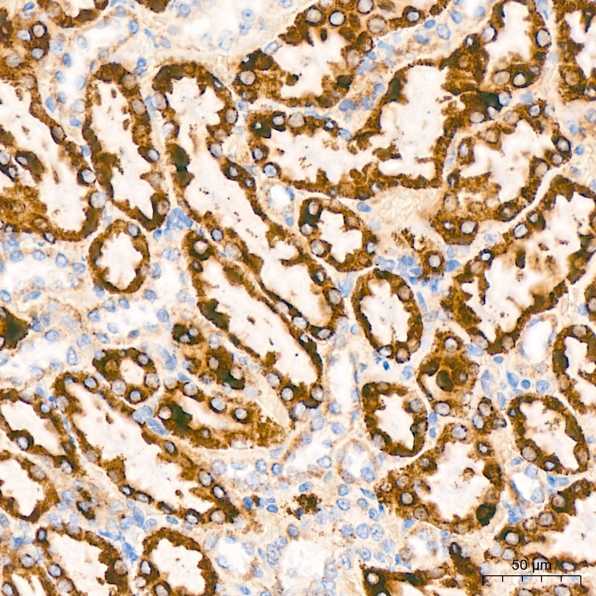
Immunohistochemistry analysis of paraffin-embedded Rat kidney tissue using TIMM50 Rabbit mAb (CAB1536) at a dilution of 1:200 (40x lens). High pressure antigen retrieval was performed with 0.01 M Tris-EDTA buffer (pH 9.0) prior to IHC staining.
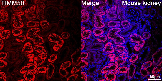
Confocal imaging of paraffin-embedded Mouse kidney tissue using TIMM50 Rabbit mAb (CAB1536, dilution 1:100) followed by a further incubation with Cy3 Goat Anti-Rabbit IgG (H+L) (CABS007, dilution 1:500) (Red). DAPI was used for nuclear staining (Blue). Objective: 40x. Perform high pressure antigen retrieval with 0.01 M citrate buffer (pH 6.0) prior to IF staining.
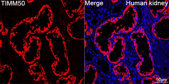
Confocal imaging of paraffin-embedded Human kidney tissue using TIMM50 Rabbit mAb (CAB1536, dilution 1:100) followed by a further incubation with Cy3 Goat Anti-Rabbit IgG (H+L) (CABS007, dilution 1:500) (Red). DAPI was used for nuclear staining (Blue). Objective: 40x. Perform high pressure antigen retrieval with 0.01 M citrate buffer (pH 6.0) prior to IF staining.