The TMOD2 Monoclonal Antibody (CAB19770) is a high-quality antibody developed for reliable detection and analysis of target proteins. This antibody is produced in rabbits and is highly specific for detecting TMOD2 in human samples, making it ideal for use in Western blot applications. By binding to the TMOD2 protein, this antibody allows for the precise detection and analysis of TMOD2 expression in various cell types.TMOD2 plays a crucial role in maintaining the structure and function of the cytoskeleton, particularly in muscle cells. Dysregulation of TMOD2 has been implicated in various diseases, including muscular dystrophy and cancer.
This antibody is validated for use in WB, IHC-P, ELISA, IF-P applications and has demonstrated reactivity against Mouse, Rat samples.
Product Name:
TMOD2 Monoclonal Antibody
SKU:
CAB19770
Size:
20μL, 100μL
Reactivity:
Mouse, Rat
Clone Number:
ARC2318
Conjugate:
Unconjugated
Immunogen:
Synthetic peptide. This information is considered to be commercially sensitive.
Recommended starting concentration is 1 μg/mL. Please optimize the concentration based on your specific assay requirements.
Synonyms:
NTMOD, N-TMOD, TMOD2
Positive Sample:
Mouse testis, Mouse brain, Rat brain
Cellular Localization:
Cytoskeleton, Nucleus, Cytoplasm, Cytoskeleton.
Calculated MW:
40kDa
Observed MW:
40kDa
This gene encodes a neuronal-specific member of the tropomodulin family of actin-regulatory proteins. The encoded protein caps the pointed end of actin filaments preventing both elongation and depolymerization. The capping activity of this protein is dependent on its association with tropomyosin. Alternatively spliced transcript variants encoding different isoforms have been described.
Purification Method
Affinity purification
Gene ID
29767
Buffer Information
Store at -20℃. Avoid freeze / thaw cycles. Buffer: PBS containing 50% glycerol and 0.05% BSA, preserved with proclin300 or sodium azide, pH 7.3.
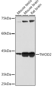
Western blot analysis of various lysates using TMOD2 Rabbit mAb (CAB19770) at 1:1000 dilution. Secondary antibody: HRP-conjugated Goat anti-Rabbit IgG (H+L) (CABS014) at 1:10000 dilution. Lysates/proteins: 25μg per lane. Blocking buffer: 3% nonfat dry milk in TBST. Detection: ECL Basic Kit (AbGn00020). Exposure time: 60s.
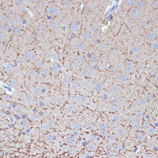
Immunohistochemistry analysis of paraffin-embedded Rat brain using TMOD2 Rabbit mAb (CAB19770) at dilution of 1:100 (40x lens). Microwave antigen retrieval performed with 0.01M Tris/EDTA Buffer (pH 9.0) prior to IHC staining.
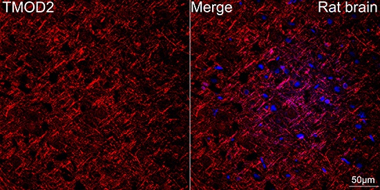
Confocal imaging of paraffin-embedded Rat brain tissue using TMOD2 Rabbit mAb (CAB19770, dilution 1:100) followed by a further incubation with Cy3 Goat Anti-Rabbit IgG (H+L) (CABS007, dilution 1:500) (Red). DAPI was used for nuclear staining (Blue). Objective: 40x. Perform microwave antigen retrieval with 0.01 M citrate buffer (pH 6.0) prior to IF staining.