The USP39 Monoclonal Antibody (CAB2389) is a high-quality antibody developed for reliable detection and analysis of target proteins. This antibody, raised in rabbits, is highly specific for USP39 and is validated for use in various applications such as Western blotting and immunofluorescence.USP39 is known to be essential for cell survival and proliferation, making it a promising target for studying cancer development and progression.
This antibody is validated for use in WB, IHC-P, IF/ICC, ELISA applications and has demonstrated reactivity against Human, Mouse, Rat samples.
Product Name:
USP39 Monoclonal Antibody
SKU:
CAB2389
Size:
20μL, 100μL
Reactivity:
Human, Mouse, Rat
Clone Number:
ARC2588
Conjugate:
Unconjugated
Immunogen:
Synthetic peptide. This information is considered to be commercially sensitive.
Recommended starting concentration is 1 μg/mL. Please optimize the concentration based on your specific assay requirements.
Synonyms:
65K, SAD1, CGI-21, HSPC332, SNRNP65, USP39
Positive Sample:
Mouse liver
Cellular Localization:
Nucleus.
Calculated MW:
65kDa
Observed MW:
65kDa
Predicted to enable thiol-dependent deubiquitinase and zinc ion binding activity. Involved in spliceosomal complex assembly. Located in nucleoplasm. Part of U4/U6 x U5 tri-snRNP complex.
Purification Method
Affinity purification
Gene ID
10713
Buffer Information
Store at -20℃. Avoid freeze / thaw cycles. Buffer: PBS containing 50% glycerol and 0.05% BSA, preserved with proclin300 or sodium azide, pH 7.3.
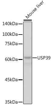
Western blot analysis of lysates from mouse liver, using USP39 Rabbit mAb (CAB2389) at 1:1000 dilution. Secondary antibody: HRP-conjugated Goat anti-Rabbit IgG (H+L) (CABS014) at 1:10000 dilution. Lysates/proteins: 25μg per lane. Blocking buffer: 3% nonfat dry milk in TBST. Detection: ECL Basic Kit (AbGn00020). Exposure time: 1s.
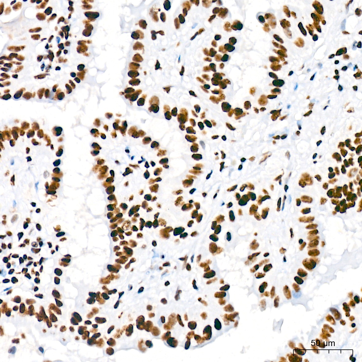
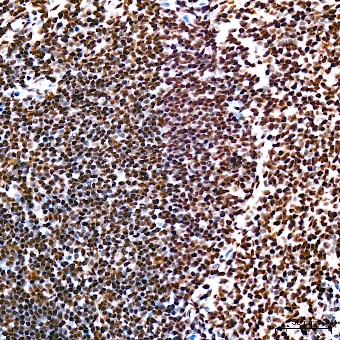
Immunohistochemistry analysis of paraffin-embedded Human spleen tissue using USP39 Rabbit mAb (CAB2389) at a dilution of 1:200 (40x lens). High pressure antigen retrieval was performed with 0.01 M citrate buffer (pH 6.0) prior to IHC staining.
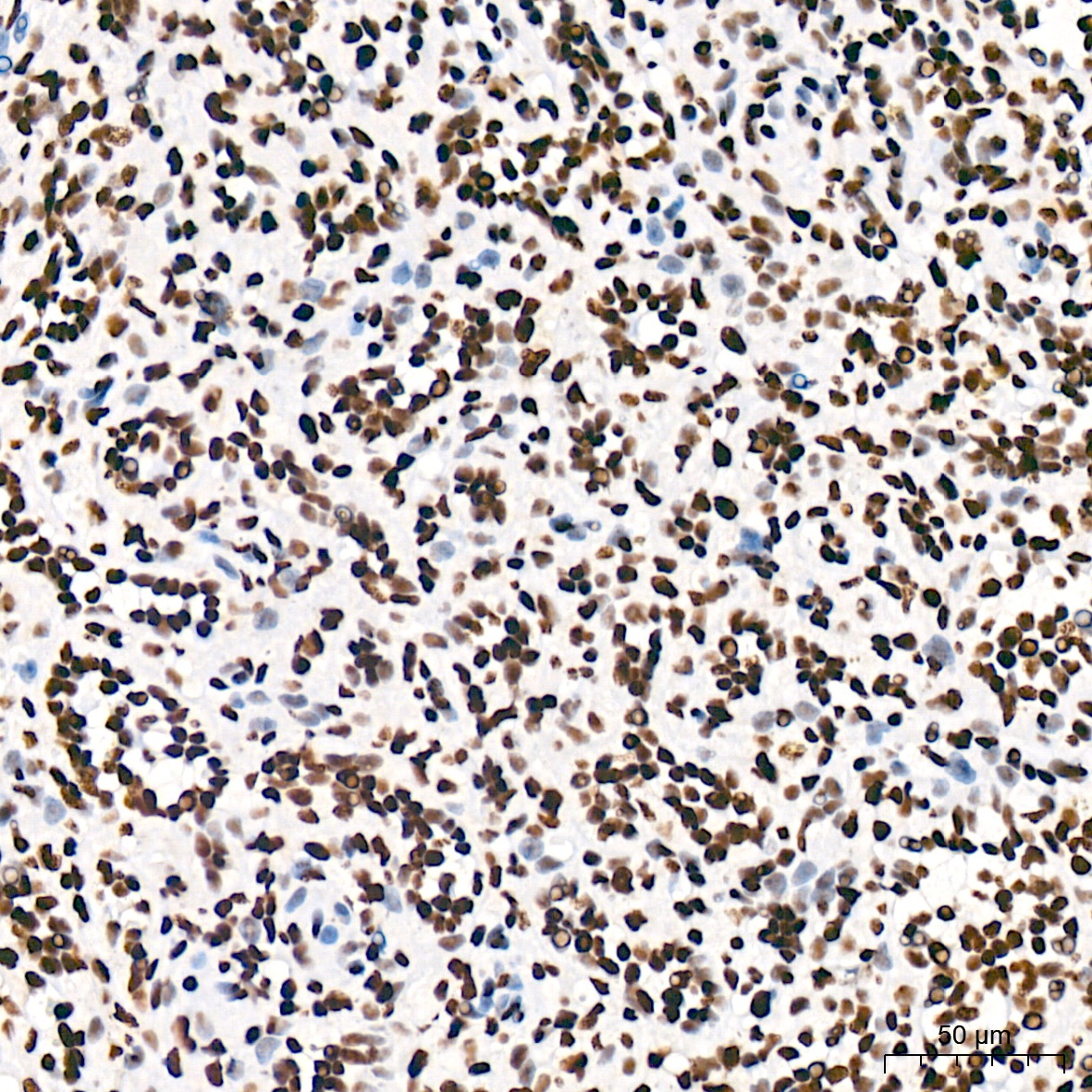
Immunohistochemistry analysis of paraffin-embedded Human thyroid cancer tissue using USP39 Rabbit mAb (CAB2389) at a dilution of 1:200 (40x lens). High pressure antigen retrieval was performed with 0.01 M citrate buffer (pH 6.0) prior to IHC staining.
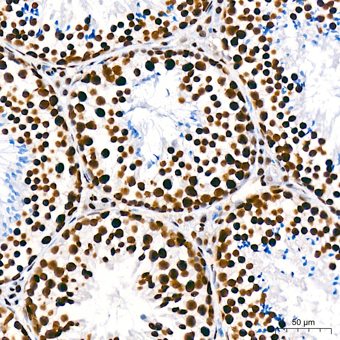
Immunohistochemistry analysis of paraffin-embedded Mouse testis tissue using USP39 Rabbit mAb (CAB2389) at a dilution of 1:200 (40x lens). High pressure antigen retrieval was performed with 0.01 M citrate buffer (pH 6.0) prior to IHC staining.
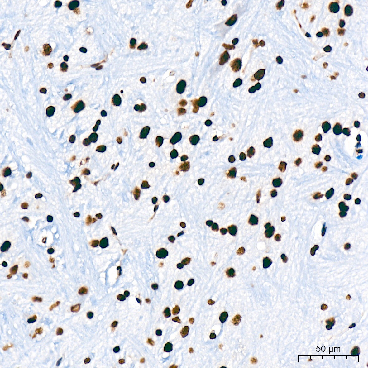
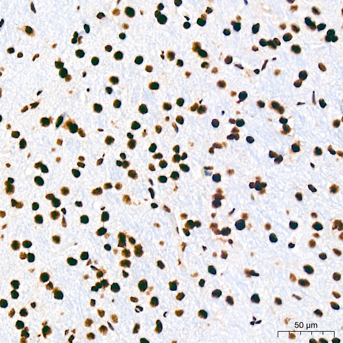
Immunohistochemistry analysis of paraffin-embedded Mouse brain tissue using USP39 Rabbit mAb (CAB2389) at a dilution of 1:200 (40x lens). High pressure antigen retrieval was performed with 0.01 M citrate buffer (pH 6.0) prior to IHC staining.
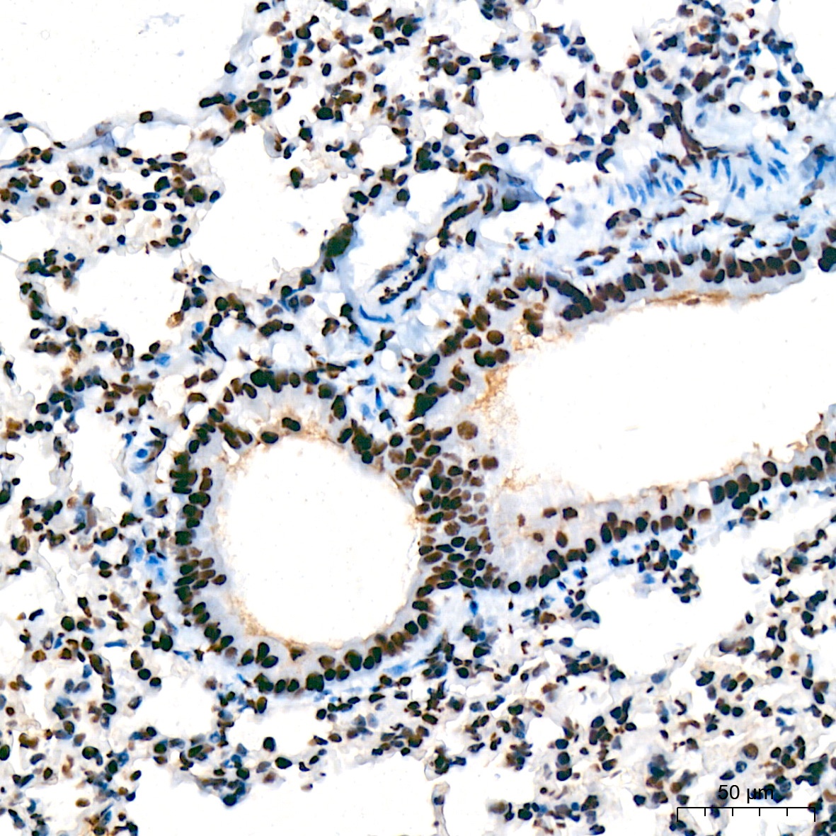
Immunohistochemistry analysis of paraffin-embedded Mouse lung tissue using USP39 Rabbit mAb (CAB2389) at a dilution of 1:200 (40x lens). High pressure antigen retrieval was performed with 0.01 M citrate buffer (pH 6.0) prior to IHC staining.
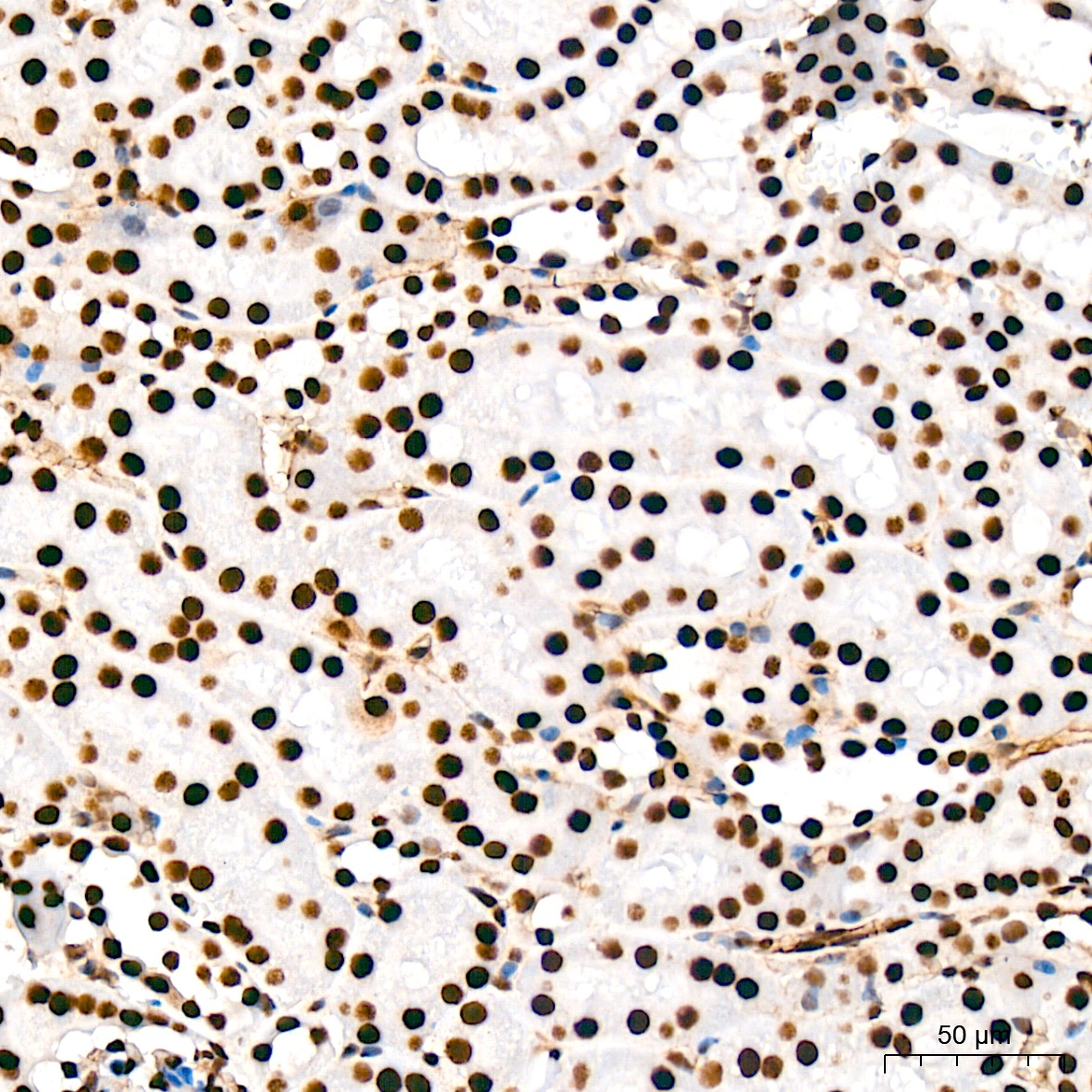
Immunohistochemistry analysis of paraffin-embedded Mouse kidney tissue using USP39 Rabbit mAb (CAB2389) at a dilution of 1:200 (40x lens). High pressure antigen retrieval was performed with 0.01 M citrate buffer (pH 6.0) prior to IHC staining.
Immunohistochemistry analysis of paraffin-embedded Mouse kidney tissue using USP39 Rabbit mAb (CAB2389) at a dilution of 1:200 (40x lens). High pressure antigen retrieval was performed with 0.01 M citrate buffer (pH 6.0) prior to IHC staining.
Immunohistochemistry analysis of paraffin-embedded Rat spleen tissue using USP39 Rabbit mAb (CAB2389) at a dilution of 1:200 (40x lens). High pressure antigen retrieval was performed with 0.01 M citrate buffer (pH 6.0) prior to IHC staining.
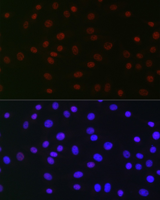
Immunofluorescence analysis of NIH/3T3 cells using USP39 Rabbit mAb (CAB2389) at dilution of 1:100 (40x lens). Secondary antibody: Cy3-conjugated Goat anti-Rabbit IgG (H+L) (CABS007) at 1:500 dilution. Blue: DAPI for nuclear staining.
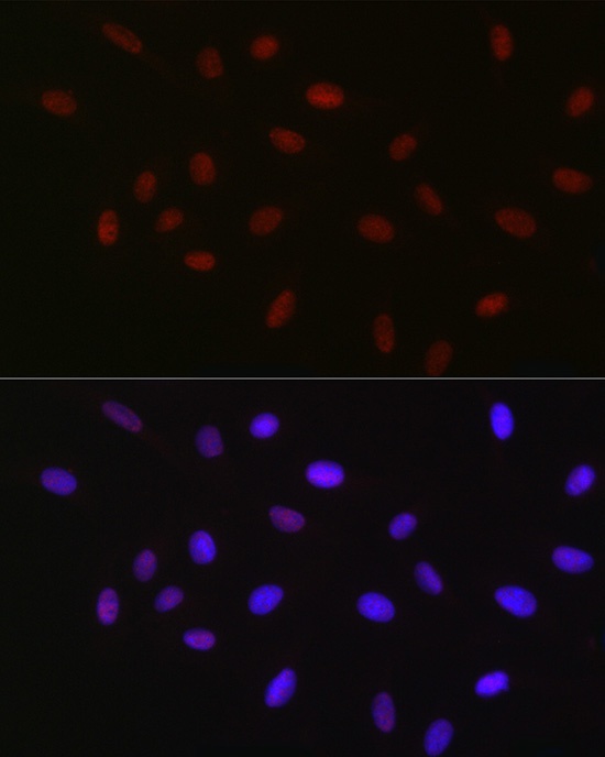
Immunofluorescence analysis of U-2 OS cells using USP39 Rabbit mAb (CAB2389) at dilution of 1:100 (40x lens). Secondary antibody: Cy3-conjugated Goat anti-Rabbit IgG (H+L) (CABS007) at 1:500 dilution. Blue: DAPI for nuclear staining.