The USP39 Monoclonal Antibody (CAB2389) is a high-quality antibody developed for reliable detection and analysis of target proteins. Predicted to enable thiol-dependent deubiquitinase and zinc ion binding activity. Involved in spliceosomal complex assembly. Located in nucleoplasm. Part of U4/U6 x U5 tri-snRNP complex. RRID Gene ID 10713 Swiss Prot Synonym 65K; SAD1; CGI-21; HSPC332; SNRNP65; USP39
This antibody is validated for use in WB, IHC-P, IF/ICC, ELISA applications and has demonstrated reactivity against Human, Mouse, Rat samples.
Product Name:
USP39 Monoclonal Antibody
SKU:
CAB2389
Size:
100μL, 20μL
Reactivity:
Human, Mouse, Rat
Clone Number:
ARC2588
Conjugate:
Unconjugated
Immunogen:
Synthetic peptide. This information is considered to be commercially sensitive.
Tested Applications:
WBIHC-PIF/ICCELISA
Recommended Dilution:
WB
1:500 - 1:1000
IHC-P
1:50 - 1:200
IF
/
ICC
1:50 - 1:200
ELISA
Recommended starting concentration is 1 μg/mL. Please optimize the concentration based on your specific assay requirements.
Synonyms:
65K, SAD1, CGI-21, HSPC332, SNRNP65, USP39
Positive Sample:
Mouse liver
Cellular Localization:
Nucleus.
Calculated MW:
65kDa
Observed MW:
65kDa
Predicted to enable thiol-dependent deubiquitinase and zinc ion binding activity. Involved in spliceosomal complex assembly. Located in nucleoplasm. Part of U4/U6 x U5 tri-snRNP complex. RRID Gene ID 10713 Swiss Prot Synonym 65K; SAD1; CGI-21; HSPC332; SNRNP65; USP39
Purification Method:
Affinity purification
Gene ID:
10713
RRID:
-
Buffer Information:
Store at -20℃. Avoid freeze / thaw cycles. Buffer: PBS containing 50% glycerol and 0.05% BSA, preserved with proclin300 or sodium azide, pH 7.3.
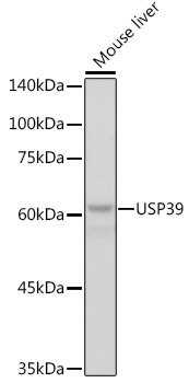
Western blot analysis of lysates from mouse liver, using USP39 Rabbit mAb (CAB2389) at 1:1000 dilution. Secondary antibody: HRP-conjugated Goat anti-Rabbit IgG (H+L) (AS014) at 1:10000 dilution. Lysates/proteins: 25μg per lane. Blocking buffer: 3% nonfat dry milk in TBST. Detection: ECL Basic Kit (AbGn00020). Exposure time: 1s.
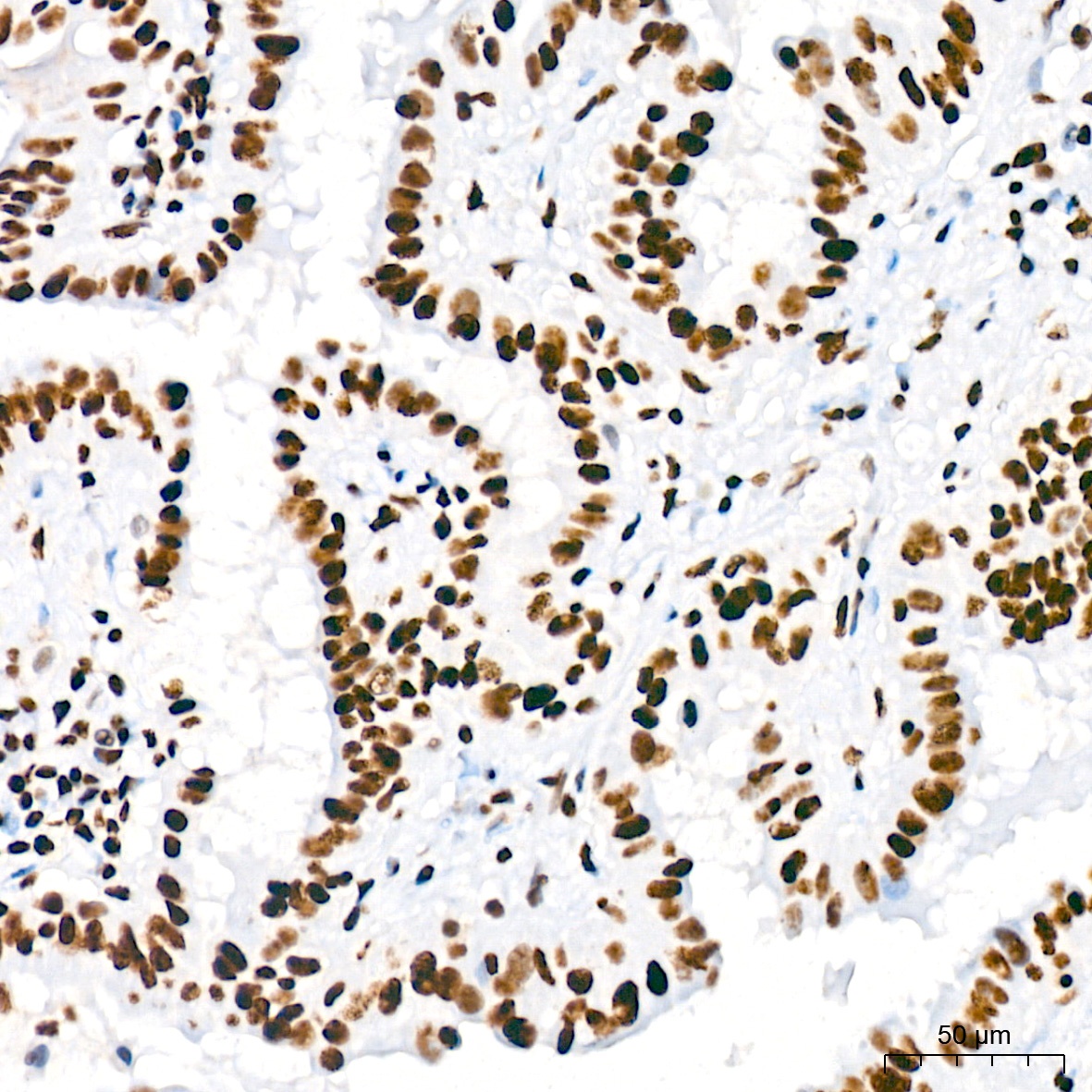
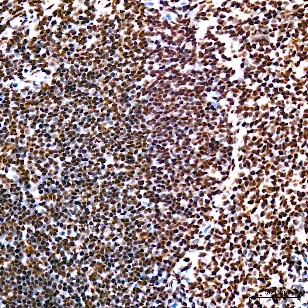
Immunohistochemistry analysis of paraffin-embedded Human spleen tissue using USP39 Rabbit mAb (CAB2389) at a dilution of 1:200 (40x lens). High pressure antigen retrieval was performed with 0.01 M citrate buffer (pH 6.0) prior to IHC staining.
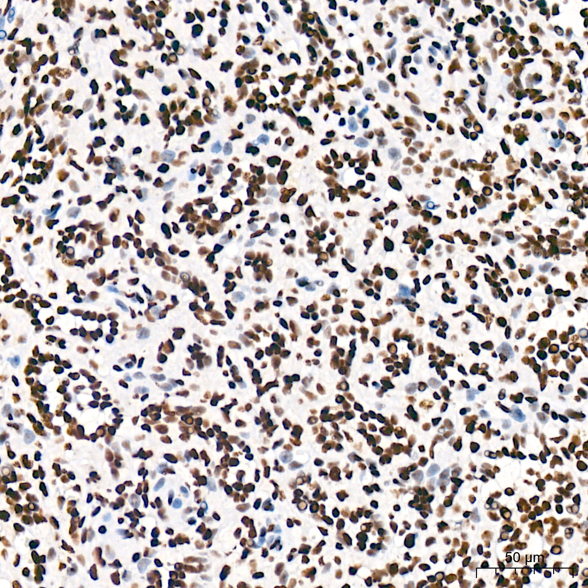
Immunohistochemistry analysis of paraffin-embedded Human thyroid cancer tissue using USP39 Rabbit mAb (CAB2389) at a dilution of 1:200 (40x lens). High pressure antigen retrieval was performed with 0.01 M citrate buffer (pH 6.0) prior to IHC staining.
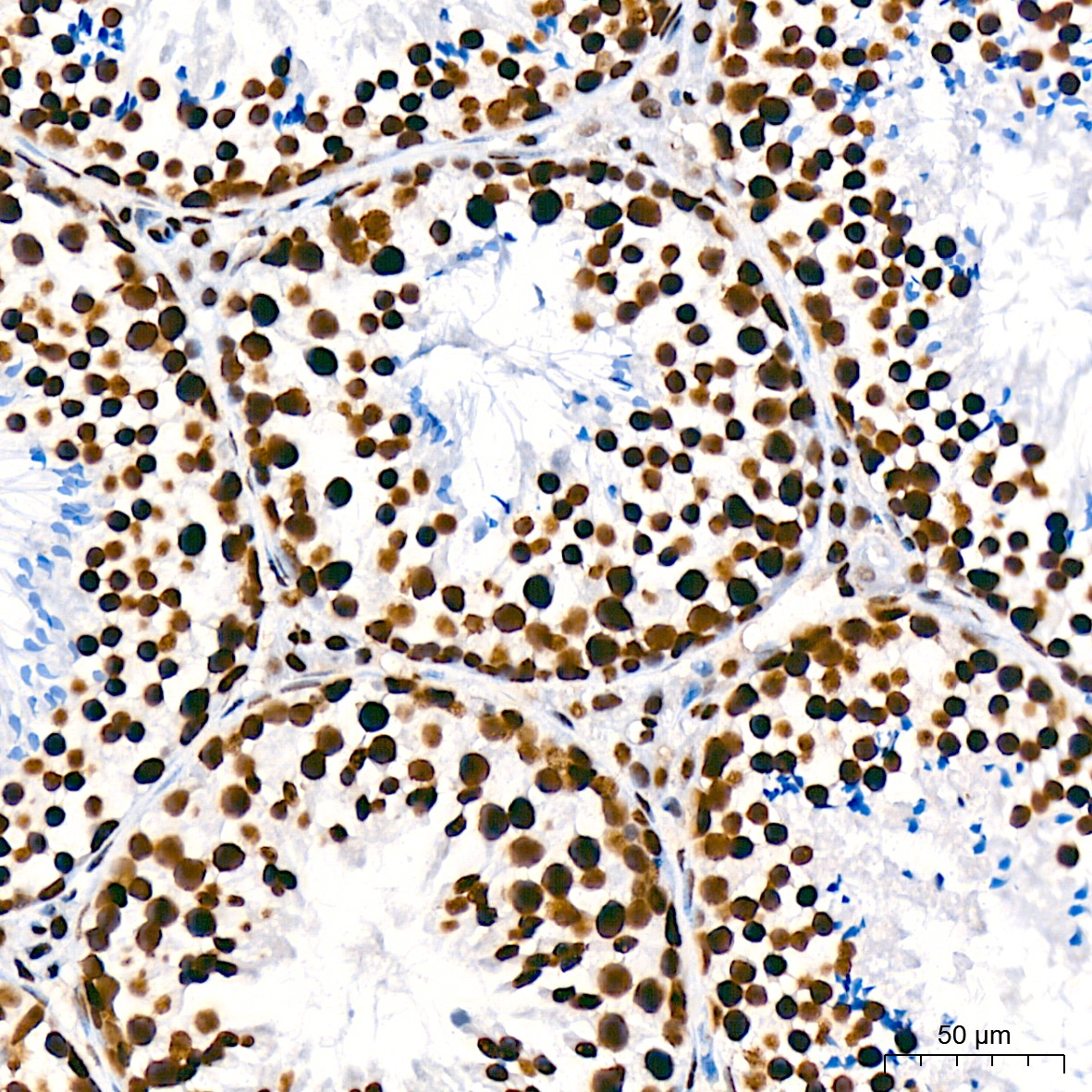
Immunohistochemistry analysis of paraffin-embedded Mouse testis tissue using USP39 Rabbit mAb (CAB2389) at a dilution of 1:200 (40x lens). High pressure antigen retrieval was performed with 0.01 M citrate buffer (pH 6.0) prior to IHC staining.
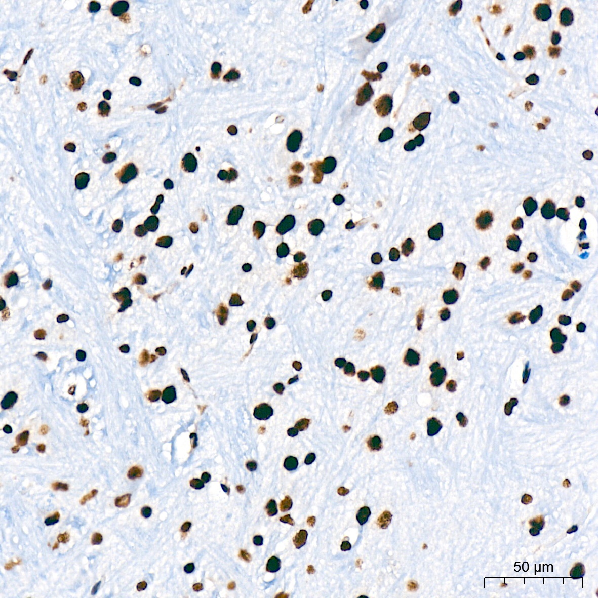
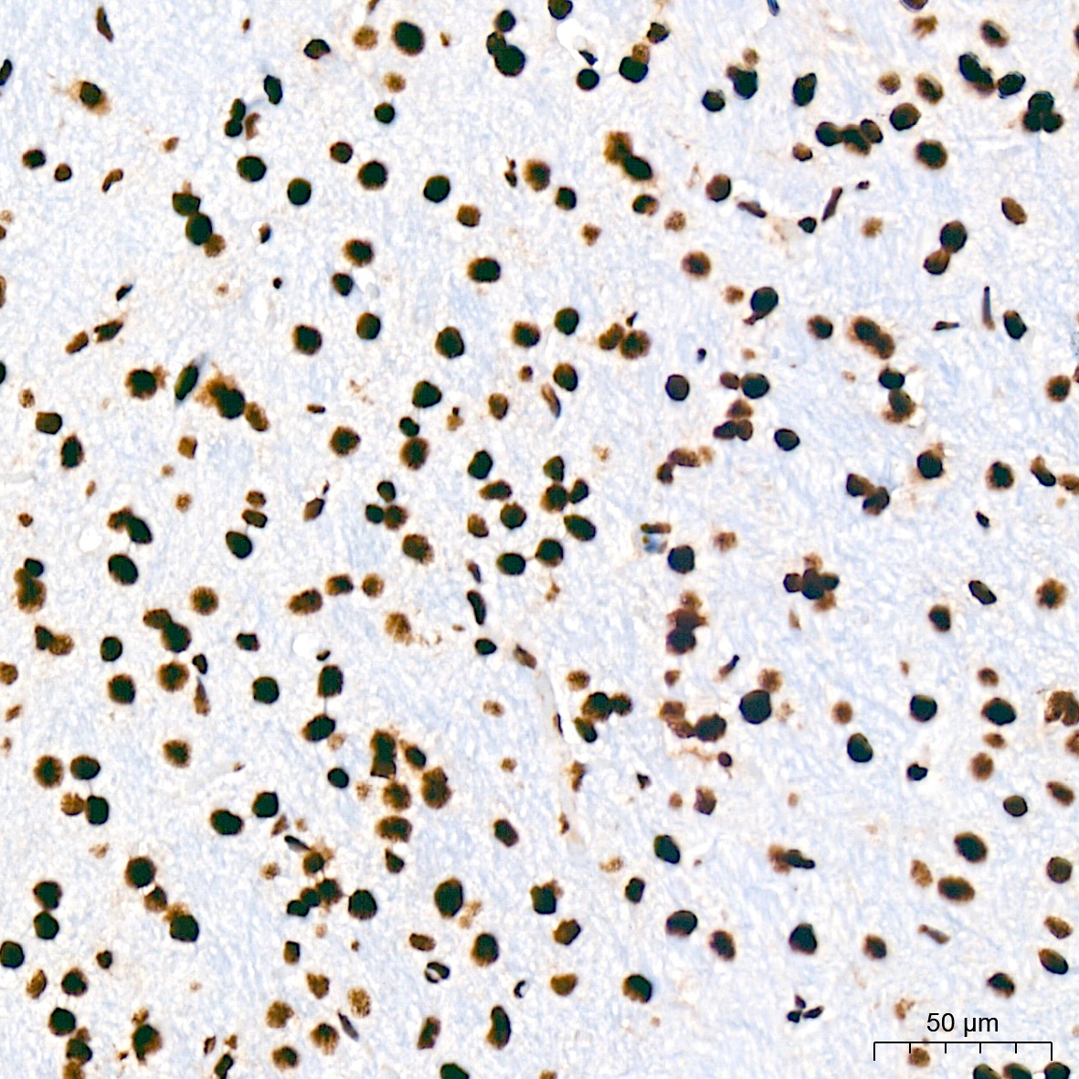
Immunohistochemistry analysis of paraffin-embedded Mouse brain tissue using USP39 Rabbit mAb (CAB2389) at a dilution of 1:200 (40x lens). High pressure antigen retrieval was performed with 0.01 M citrate buffer (pH 6.0) prior to IHC staining.
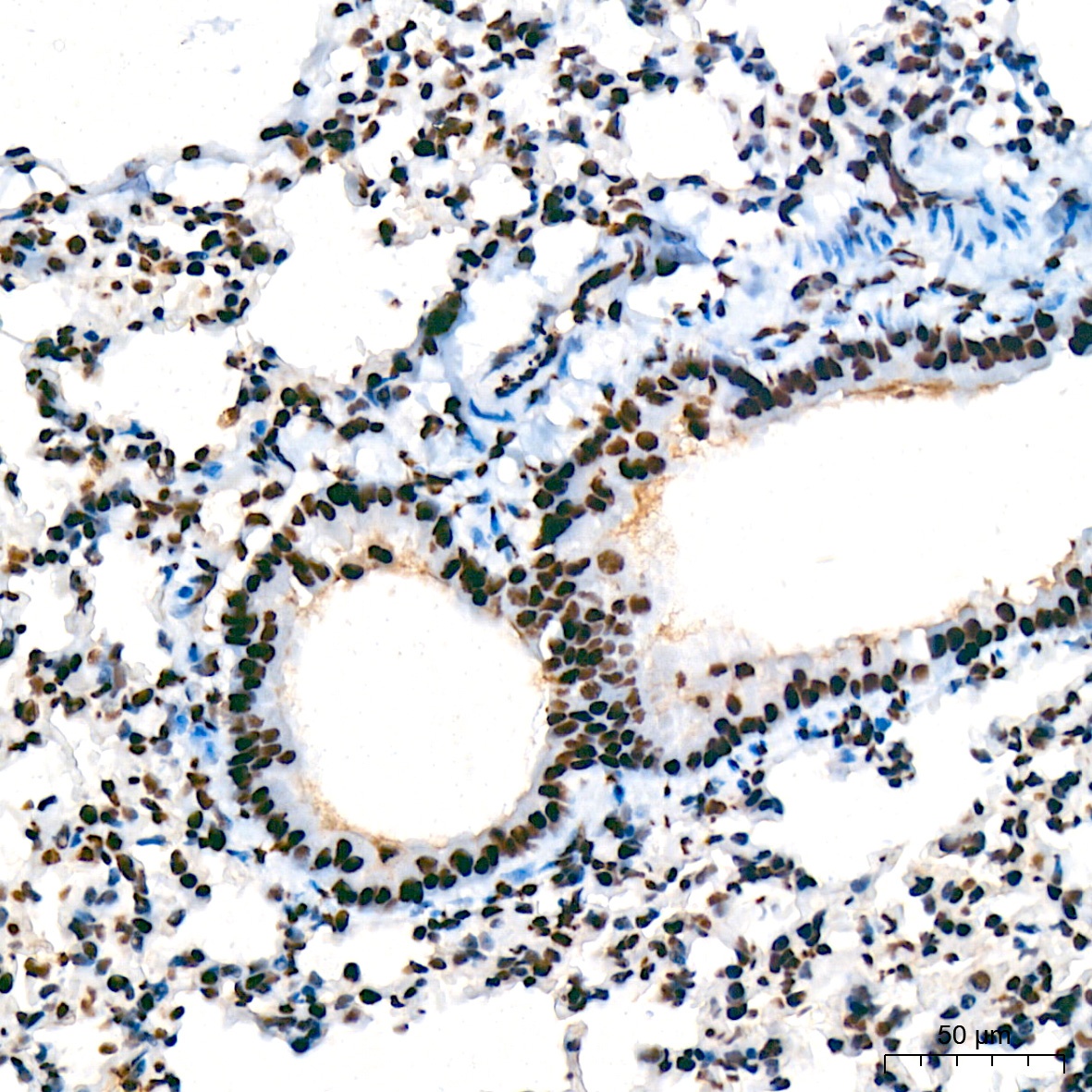
Immunohistochemistry analysis of paraffin-embedded Mouse lung tissue using USP39 Rabbit mAb (CAB2389) at a dilution of 1:200 (40x lens). High pressure antigen retrieval was performed with 0.01 M citrate buffer (pH 6.0) prior to IHC staining.
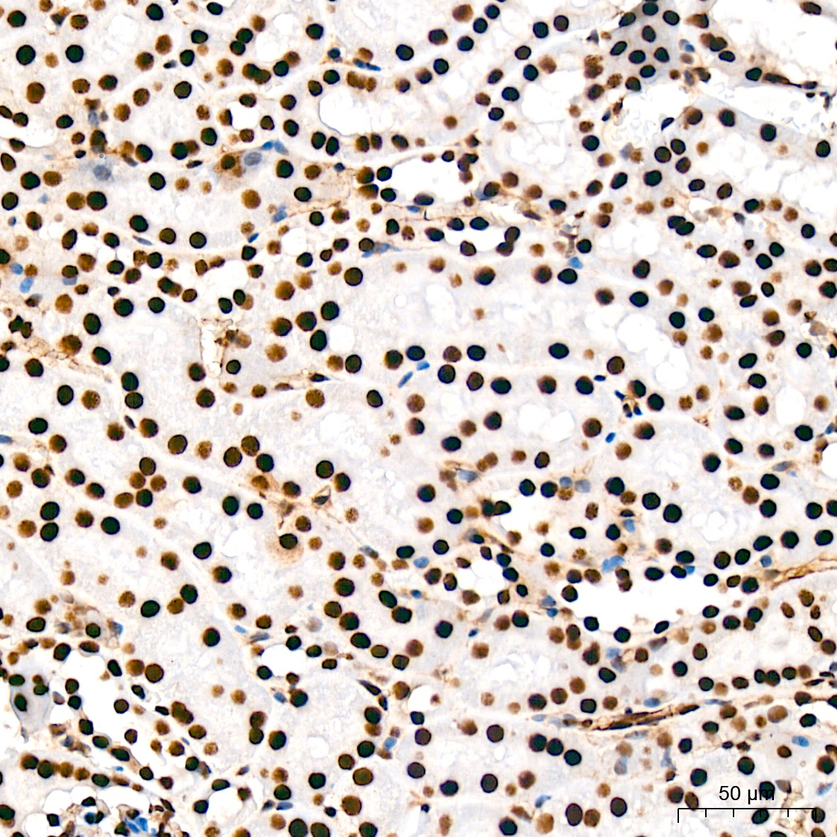
Immunohistochemistry analysis of paraffin-embedded Mouse kidney tissue using USP39 Rabbit mAb (CAB2389) at a dilution of 1:200 (40x lens). High pressure antigen retrieval was performed with 0.01 M citrate buffer (pH 6.0) prior to IHC staining.
Immunohistochemistry analysis of paraffin-embedded Mouse kidney tissue using USP39 Rabbit mAb (CAB2389) at a dilution of 1:200 (40x lens). High pressure antigen retrieval was performed with 0.01 M citrate buffer (pH 6.0) prior to IHC staining.
Immunohistochemistry analysis of paraffin-embedded Rat spleen tissue using USP39 Rabbit mAb (CAB2389) at a dilution of 1:200 (40x lens). High pressure antigen retrieval was performed with 0.01 M citrate buffer (pH 6.0) prior to IHC staining.
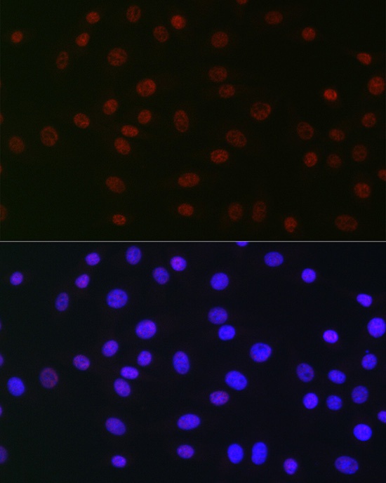
Immunofluorescence analysis of NIH/3T3 cells using USP39 Rabbit mAb (CAB2389) at dilution of 1:100 (40x lens). Secondary antibody: Cy3-conjugated Goat anti-Rabbit IgG (H+L) (AS007) at 1:500 dilution. Blue: DAPI for nuclear staining.
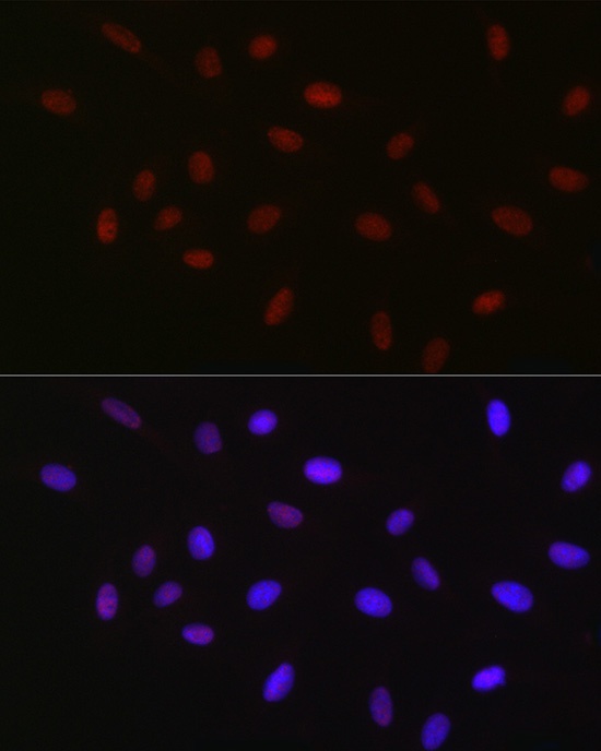
Immunofluorescence analysis of U-2 OS cells using USP39 Rabbit mAb (CAB2389) at dilution of 1:100 (40x lens). Secondary antibody: Cy3-conjugated Goat anti-Rabbit IgG (H+L) (AS007) at 1:500 dilution. Blue: DAPI for nuclear staining.