The XPD/ERCC2 Monoclonal Antibody (CAB19241) is a high-quality antibody developed for reliable detection and analysis of target proteins. This antibody, produced in rabbits, exhibits high reactivity with human samples and is specifically validated for use in Western blot applications. By targeting the XPD/ERCC2 protein, researchers can explore its functions in DNA repair pathways and its implications in various cellular processes.XPD/ERCC2, a member of the nucleotide excision repair complex, is essential for maintaining genomic stability and preventing mutations that can lead to diseases such as cancer.
This antibody is validated for use in WB, IHC-P, ELISA applications and has demonstrated reactivity against Human, Mouse, Rat samples.
Product Name:
XPD/ERCC2 Monoclonal Antibody
SKU:
CAB19241
Size:
20μL, 100μL
Reactivity:
Human, Mouse, Rat
Clone Number:
ARC2401
Conjugate:
Unconjugated
Immunogen:
Synthetic peptide. This information is considered to be commercially sensitive.
Recommended starting concentration is 1 μg/mL. Please optimize the concentration based on your specific assay requirements.
Synonyms:
EM9, TTD, XPD, TTD1, COFS2, TFIIH, XPD/ERCC2
Positive Sample:
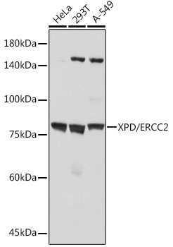
HeLa, 293T, A-549
Cellular Localization:
Cytoplasm, Nucleus, Cytoskeleton, Spindle.
Calculated MW:
87kDa
Observed MW:
80kDa
The nucleotide excision repair pathway is a mechanism to repair damage to DNA. The protein encoded by this gene is involved in transcription-coupled nucleotide excision repair and is an integral member of the basal transcription factor BTF2/TFIIH complex. The gene product has ATP-dependent DNA helicase activity and belongs to the RAD3/XPD subfamily of helicases. Defects in this gene can result in three different disorders, the cancer-prone syndrome xeroderma pigmentosum complementation group D, trichothiodystrophy, and Cockayne syndrome. Alternatively spliced transcript variants encoding different isoforms have been found for this gene.
Purification Method
Affinity purification
Gene ID
2068
Buffer Information
Store at -20℃. Avoid freeze / thaw cycles. Buffer: PBS containing 50% glycerol and 0.05% BSA, preserved with proclin300 or sodium azide, pH 7.3.
Western blot analysis of various lysates using XPD/ERCC2 Rabbit mAb (CAB19241) at 1:1000 dilution. Secondary antibody: HRP-conjugated Goat anti-Rabbit IgG (H+L) (CABS014) at 1:10000 dilution. Lysates/proteins: 25μg per lane. Blocking buffer: 3% nonfat dry milk in TBST. Detection: ECL Basic Kit (AbGn00020). Exposure time: 10s.
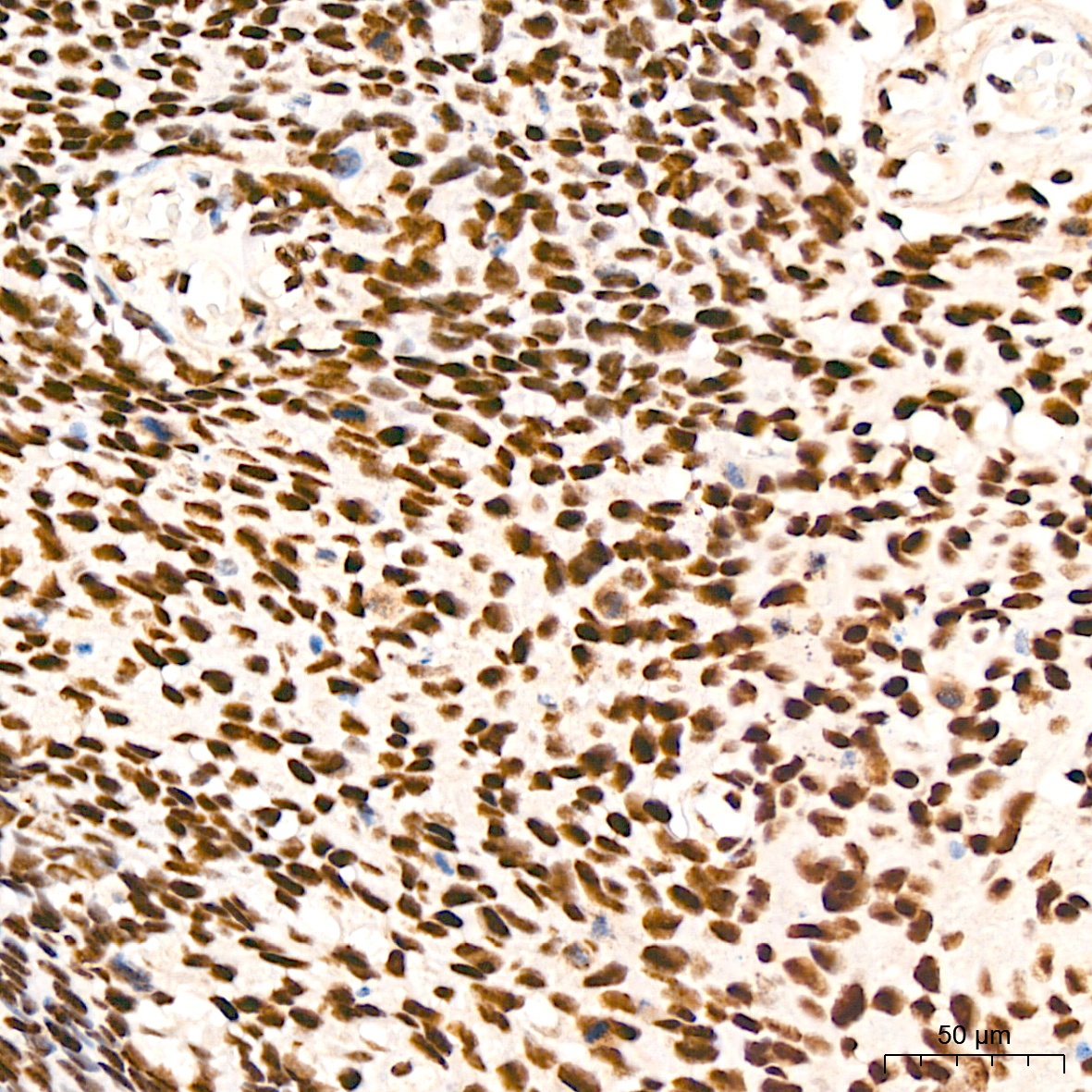
Immunohistochemistry analysis of paraffin-embedded Human cervix cancer tissue using XPD/ERCC2 Rabbit mAb (CAB19241) at a dilution of 1:200 (40x lens). High pressure antigen retrieval was performed with 0.01 M Tris-EDTA buffer (pH 9.0) prior to IHC staining.
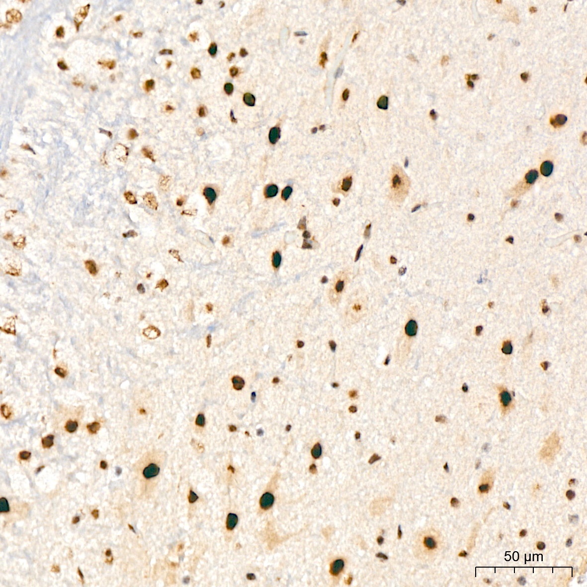
Immunohistochemistry analysis of paraffin-embedded Mouse brain tissue using XPD/ERCC2 Rabbit mAb (CAB19241) at a dilution of 1:200 (40x lens). High pressure antigen retrieval was performed with 0.01 M Tris-EDTA buffer (pH 9.0) prior to IHC staining.
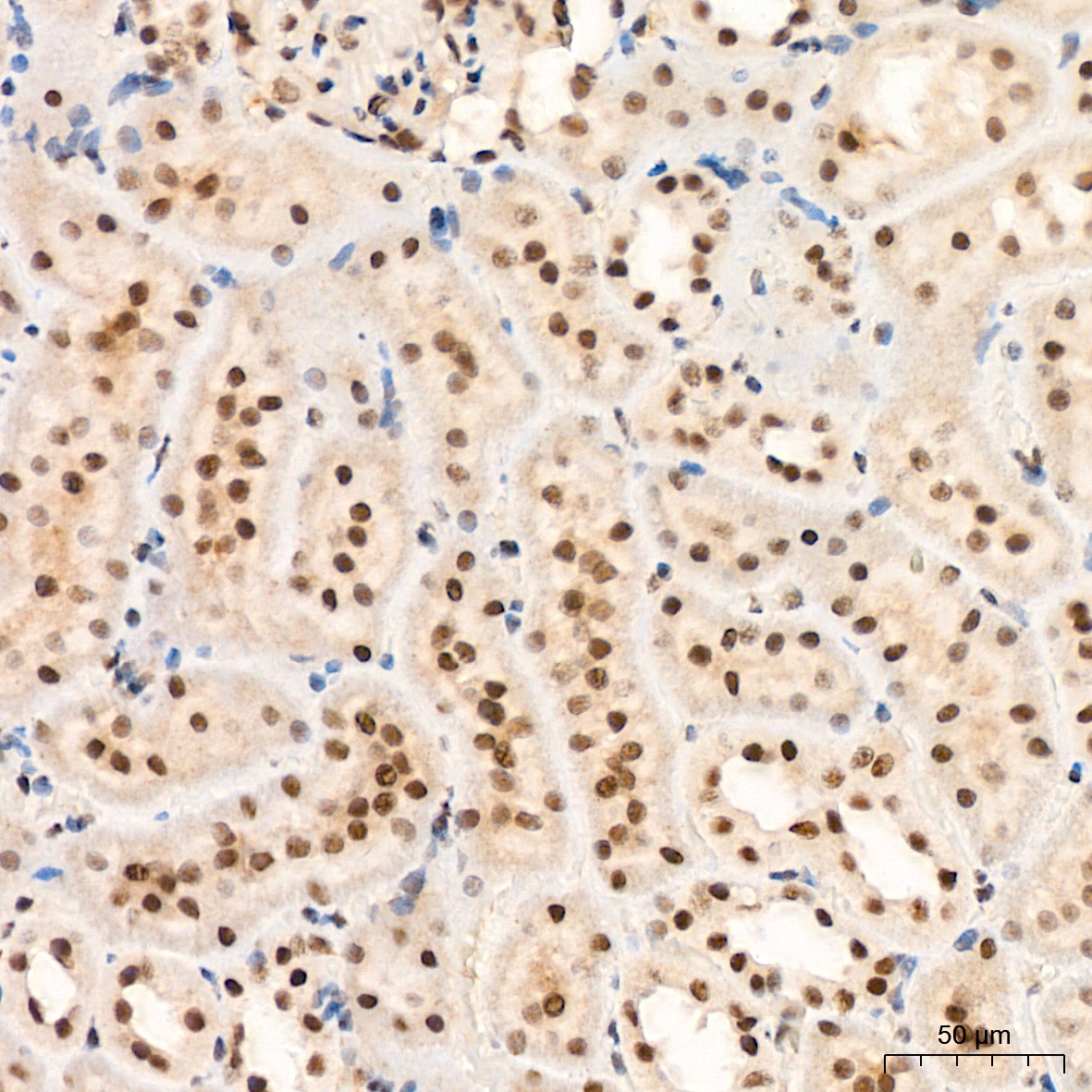
Immunohistochemistry analysis of paraffin-embedded Rat kidney tissue using XPD/ERCC2 Rabbit mAb (CAB19241) at a dilution of 1:200 (40x lens). High pressure antigen retrieval was performed with 0.01 M Tris-EDTA buffer (pH 9.0) prior to IHC staining.