The ZBTB7A/FBI-1/LRF Monoclonal Antibody (CAB19785) is a high-quality antibody developed for reliable detection and analysis of target proteins. This polyclonal antibody, raised in rabbits, is highly specific for human samples and is validated for use in Western blot and immunohistochemistry applications.ZBTB7A, also known as FBI-1 or LRF, plays a crucial role in gene expression regulation and has been implicated in various diseases such as cancer and hematological malignancies.
This antibody is validated for use in WB, IHC-P, IF/ICC, ELISA applications and has demonstrated reactivity against Human samples.
Product Name:
ZBTB7A/FBI-1/LRF Monoclonal Antibody
SKU:
CAB19785
Size:
20μL, 100μL
Reactivity:
Human
Clone Number:
ARC2320
Conjugate:
Unconjugated
Immunogen:
Synthetic peptide. This information is considered to be commercially sensitive.
Enables several functions, including SMAD binding activity; androgen receptor binding activity; and transcription corepressor binding activity. Involved in several processes, including erythrocyte maturation; negative regulation of signal transduction; and regulation of nucleobase-containing compound metabolic process. Located in cytoplasm and nucleus. Colocalizes with DNA-dependent protein kinase complex and NuRD complex.
Purification Method
Affinity purification
Gene ID
51341
Buffer Information
Store at -20℃. Avoid freeze / thaw cycles. Buffer: PBS containing 50% glycerol and 0.05% BSA, preserved with proclin300 or sodium azide, pH 7.3.
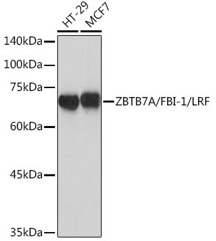
Western blot analysis of various lysates using ZBTB7A/FBI-1/LRF Rabbit mAb (CAB19785) at 1:1000 dilution. Secondary antibody: HRP-conjugated Goat anti-Rabbit IgG (H+L) (CABS014) at 1:10000 dilution. Lysates/proteins: 25μg per lane. Blocking buffer: 3% nonfat dry milk in TBST. Detection: ECL Basic Kit (AbGn00020). Exposure time: 1s.
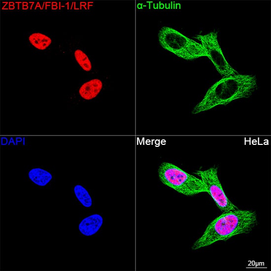
Confocal imaging of HeLa cells using ZBTB7A/FBI-1/LRF Rabbit mAb (CAB19785, dilution 1:200) followed by a further incubation with Cy3 Goat Anti-Rabbit IgG (H+L) (CABS007, dilution 1:500) (Red). The cells were counterstained with α-Tubulin Mouse mAb (AC012, dilution 1:400) followed by incubation with ABflo® 488-conjugated Goat Anti-Mouse IgG (H+L) Ab (CABS076, dilution 1:500) (Green). DAPI was used for nuclear staining (Blue). Objective: 100x.
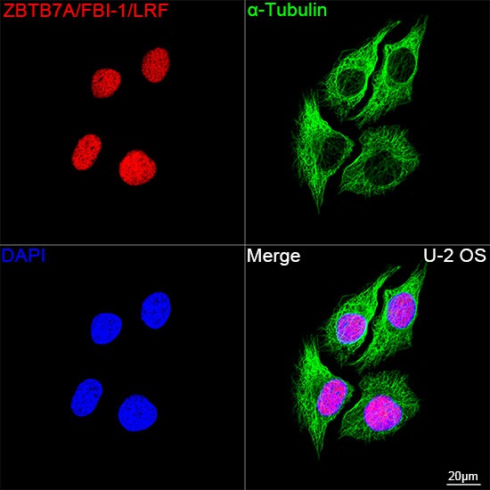
Confocal imaging of U-2 OS cells using ZBTB7A/FBI-1/LRF Rabbit mAb (CAB19785, dilution 1:200) followed by a further incubation with Cy3 Goat Anti-Rabbit IgG (H+L) (CABS007, dilution 1:500) (Red). The cells were counterstained with α-Tubulin Mouse mAb (AC012, dilution 1:400) followed by incubation with ABflo® 488-conjugated Goat Anti-Mouse IgG (H+L) Ab (CABS076, dilution 1:500) (Green). DAPI was used for nuclear staining (Blue). Objective: 100x.
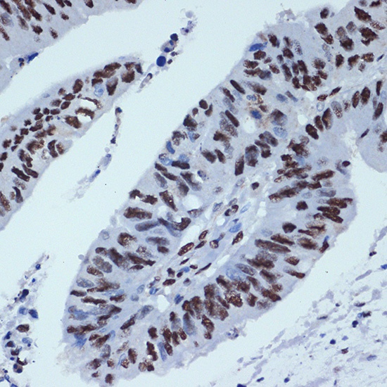
Immunohistochemistry analysis of paraffin-embedded Human colon carcinoma using ZBTB7A/FBI-1/LRF Rabbit mAb (CAB19785) at dilution of 1:100 (40x lens). Microwave antigen retrieval performed with 0.01M Tris/EDTA Buffer (pH 9.0) prior to IHC staining.