The APCS Antibody (CAB1996) is a high-quality antibody developed for reliable detection and analysis of target proteins. This antibody, raised in rabbits, is highly reactive with human samples and has been validated for use in various applications, including Western blotting.APCs are essential in the immune system as they engulf and present antigens to T cells, initiating the adaptive immune response. Studying APCs can provide valuable insights into how the immune system functions and responds to pathogens, allergens, and cancer cells.
This antibody is validated for use in WB, IHC-P, ELISA applications and has demonstrated reactivity against Human, Mouse, Rat samples.
Product Name:
APCS Antibody
SKU:
CAB1996
Size:
20μL, 100μL
Reactivity:
Human, Mouse, Rat
Conjugate:
Unconjugated
Immunogen:
Synthetic peptide. This information is considered to be commercially sensitive.
Recommended starting concentration is 1 μg/mL. Please optimize the concentration based on your specific assay requirements.
Synonyms:
SAP, PTX2, HEL-S-92n, APCS
Positive Sample:
293T, Mouse kidney, Rat liver
Cellular Localization:
Secreted.
Calculated MW:
25kDa
Observed MW:
25kDa
The protein encoded by this gene is a glycoprotein, belonging to the pentraxin family of proteins, which has a characteristic pentameric organization. These family members have considerable sequence homology which is thought to be the result of gene duplication. The binding of the encoded protein to proteins in the pathological amyloid cross-beta fold suggests its possible role as a chaperone. This protein is also thought to control the degradation of chromatin. It has been demonstrated that this protein binds to apoptotic cells at an early stage, which raises the possibility that it is involved in dealing with apoptotic cells in vivo.
Purification Method
Affinity purification
Gene ID
325
RRID
AB_2764021
Buffer Information
Store at -20℃. Avoid freeze / thaw cycles. Buffer: PBS containing 50% glycerol, preserved with proclin300 or sodium azide, pH 7.3.
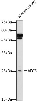
Western blot analysis of lysates from Mouse kidney, using APCS Rabbit pAb (CAB1996) at 1:1000 dilution. Secondary antibody: HRP-conjugated Goat anti-Rabbit IgG (H+L) (CABS014) at 1:10000 dilution. Lysates/proteins: 25μg per lane. Blocking buffer: 3% nonfat dry milk in TBST. Detection: ECL Basic Kit (AbGn00020). Exposure time: 3s.
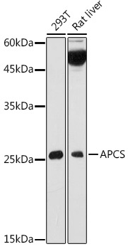
Western blot analysis of various lysates using APCS Rabbit pAb (CAB1996) at 1:1000 dilution. Secondary antibody: HRP-conjugated Goat anti-Rabbit IgG (H+L) (CABS014) at 1:10000 dilution. Lysates/proteins: 25μg per lane. Blocking buffer: 3% nonfat dry milk in TBST. Detection: ECL Basic Kit (AbGn00020). Exposure time: 10s.
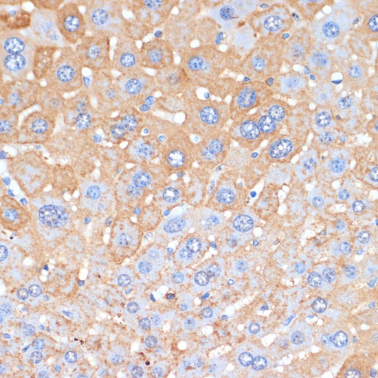
Immunohistochemistry analysis of paraffin-embedded Mouse liver using APCS Rabbit pAb (CAB1996) at dilution of 1:100 (40x lens). Microwave antigen retrieval performed with 0.01M PBS Buffer (pH 7.2) prior to IHC staining.