The APOA4 Antibody (CAB9792) is a high-quality antibody developed for reliable detection and analysis of target proteins. This antibody, raised in rabbits, is highly specific to human samples and has been validated for use in various applications, including Western blotting and immunohistochemistry.Apolipoprotein A-IV is a protein involved in the transport of dietary lipids in the bloodstream and has been shown to play a role in regulating cholesterol metabolism and inflammation. By targeting apolipoprotein A-IV with this antibody, researchers can gain insights into its function and potential implications for conditions such as cardiovascular disease and metabolic disorders.
This antibody is validated for use in WB, ELISA applications and has demonstrated reactivity against Human, Mouse, Rat samples.
Product Name:
APOA4 Antibody
SKU:
CAB9792
Size:
20μL, 100μL
Reactivity:
Human, Mouse, Rat
Conjugate:
Unconjugated
Immunogen:
Synthetic peptide. This information is considered to be commercially sensitive.
Recommended starting concentration is 1 μg/mL. Please optimize the concentration based on your specific assay requirements.
Synonyms:
APOA4
Positive Sample:
HepG2, Mouse small intestine, Rat liver
Cellular Localization:
Secreted.
Calculated MW:
45kDa
Observed MW:
45kDa
Apoliprotein (apo) A-IV gene contains 3 exons separated by two introns. A sequence polymorphism has been identified in the 3'UTR of the third exon. The primary translation product is a 396-residue preprotein which after proteolytic processing is secreted its primary site of synthesis, the intestine, in association with chylomicron particles. Although its precise function is not known, apo A-IV is a potent activator of lecithin-cholesterol acyltransferase in vitro.
Purification Method
Affinity purification
Gene ID
337
RRID
AB_2768394
Buffer Information
Store at -20℃. Avoid freeze / thaw cycles. Buffer: PBS containing 50% glycerol, preserved with proclin300 or sodium azide, pH 7.3.
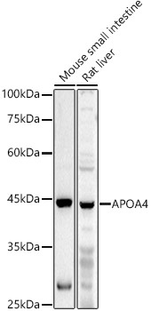
Western blot analysis of various lysates using APOA4 Rabbit pAb (CAB9792) at 1:1000 dilution. Secondary antibody: HRP-conjugated Goat anti-Rabbit IgG (H+L) (CABS014) at 1:10000 dilution. Lysates/proteins: 25μg per lane. Blocking buffer: 3% nonfat dry milk in TBST. Detection: ECL Basic Kit (AbGn00020). Exposure time: 30s.
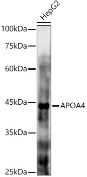
Western blot analysis of lysates from HepG2 cells, using APOA4 Rabbit pAb (CAB9792) at 1:1000 dilution. Secondary antibody: HRP-conjugated Goat anti-Rabbit IgG (H+L) (CABS014) at 1:10000 dilution. Lysates/proteins: 25μg per lane. Blocking buffer: 3% nonfat dry milk in TBST. Detection: ECL Basic Kit (AbGn00020). Exposure time: 90s.