The ApoM Monoclonal Antibody (CAB5144) is a high-quality antibody developed for reliable detection and analysis of target proteins. The protein encoded by this gene is an apolipoprotein and member of the lipocalin protein family. It is found associated with high density lipoproteins and to a lesser extent with low density lipoproteins and triglyceride-rich lipoproteins. The encoded protein is secreted through the plasma membrane but remains membrane-bound, where it is involved in lipid transport. Alternate splicing results in both coding and non-coding variants of this gene.
This antibody is validated for use in IF/ICC, ELISA, IF-P applications and has demonstrated reactivity against Human samples.
Product Name:
ApoM Monoclonal Antibody
SKU:
CAB5144
Size:
100μL, 20μL
Reactivity:
Human
Clone Number:
ARC1210
Conjugate:
Unconjugated
Immunogen:
Synthetic peptide. This information is considered to be commercially sensitive.
Tested Applications:
IF/ICCELISAIF-P
Recommended Dilution:
IF/ICC
1:50 - 1:200
IF-P
1:50 - 1:200
ELISA
Recommended starting concentration is 1 μg/mL. Please optimize the concentration based on your specific assay requirements.
Synonyms:
G3a, NG20, apo-M, HSPC336, ApoM
Cellular Localization:
Secreted.
Calculated MW:
21kDa
Observed MW:
Refer to figures
The protein encoded by this gene is an apolipoprotein and member of the lipocalin protein family. It is found associated with high density lipoproteins and to a lesser extent with low density lipoproteins and triglyceride-rich lipoproteins. The encoded protein is secreted through the plasma membrane but remains membrane-bound, where it is involved in lipid transport. Alternate splicing results in both coding and non-coding variants of this gene.
Purification Method
Affinity purification
Gene ID
55937
RRID
AB_2863465
Buffer Information
Store at -20℃. Avoid freeze / thaw cycles. Buffer: PBS containing 50% glycerol and 0.05% BSA, preserved with proclin300 or sodium azide, pH 7.3.
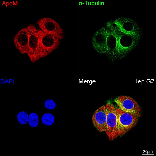
Confocal imaging of Hep G2 cells using ApoM Rabbit mAb (CAB5144, dilution 1:100) followed by a further incubation with Cy3 Goat Anti-Rabbit IgG (H+L) (AS007, dilution 1:500) (Red). The cells were counterstained with α-Tubulin Mouse mAb (AC012, dilution 1:400) followed by incubation with ABflo® 488-conjugated Goat Anti-Mouse IgG (H+L) Ab (AS076, dilution 1:500) (Green). DAPI was used for nuclear staining (Blue). Objective: 100x.
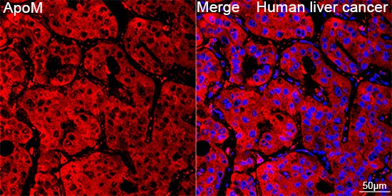
Confocal imaging of paraffin-embedded Human liver cancer tissue using ApoM Rabbit mAb (CAB5144, dilution 1:100) followed by a further incubation with Cy3 Goat Anti-Rabbit IgG (H+L) (AS007, dilution 1:500) (Red). DAPI was used for nuclear staining (Blue). Objective: 40x. Perform high pressure antigen retrieval with 0.01 M citrate buffer (pH 6.0) prior to IF staining.