The Aquaporin-1 (AQP1) Monoclonal Antibody (CAB4195) is a high-quality antibody developed for reliable detection and analysis of target proteins. This antibody, produced using rabbit monoclonal technology, is highly specific and sensitive for detecting AQP1 in human samples.AQP1 is a key player in various physiological processes, including fluid balance, urine concentration, and cell migration. Dysregulation of AQP1 has been implicated in numerous diseases, such as kidney disorders, brain edema, and cancer.
This antibody is validated for use in WB, IHC-P, ELISA, IF-P applications and has demonstrated reactivity against Human, Mouse, Rat samples.
Product Name:
Aquaporin-1 (AQP1) Monoclonal Antibody
SKU:
CAB4195
Size:
20μL, 100μL
Reactivity:
Human, Mouse, Rat
Clone Number:
ARC0925
Conjugate:
Unconjugated
Immunogen:
Synthetic peptide. This information is considered to be commercially sensitive.
Recommended starting concentration is 1 μg/mL. Please optimize the concentration based on your specific assay requirements.
Synonyms:
CO, CHIP28, AQP-CHIP, Aquaporin-1 (AQP1)
Positive Sample:
Mouse liver, Rat spleen, Rat kidney
Cellular Localization:
Cell Membrane, Multi-Pass Membrane Protein.
Calculated MW:
29kDa
Observed MW:
29kDa
This gene encodes a small integral membrane protein with six bilayer spanning domains that functions as a water channel protein. This protein permits passive transport of water along an osmotic gradient. This gene is a possible candidate for disorders involving imbalance in ocular fluid movement.
Purification Method
Affinity purification
Gene ID
358
RRID
AB_2863207
Buffer Information
Store at -20℃. Avoid freeze / thaw cycles. Buffer: PBS containing 50% glycerol and 0.05% BSA, preserved with proclin300 or sodium azide, pH 7.3.
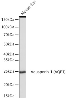
Western blot analysis of lysates from Mouse liver, using Aquaporin-1 (Aquaporin-1 (AQP1)) Rabbit mAb (CAB4195) at 1:1000 dilution. Secondary antibody: HRP-conjugated Goat anti-Rabbit IgG (H+L) (CABS014) at 1:10000 dilution. Lysates/proteins: 25μg per lane. Blocking buffer: 3% nonfat dry milk in TBST. Detection: ECL Basic Kit (AbGn00020). Exposure time: 1s.
Western blot analysis of various lysates using Aquaporin-1 (Aquaporin-1 (AQP1)) Rabbit mAb (CAB4195) at 1:1000 dilution. Secondary antibody: HRP-conjugated Goat anti-Rabbit IgG (H+L) (CABS014) at 1:10000 dilution. Lysates/proteins: 25μg per lane. Blocking buffer: 3% nonfat dry milk in TBST. Detection: ECL Basic Kit (AbGn00020). Exposure time: 10s.
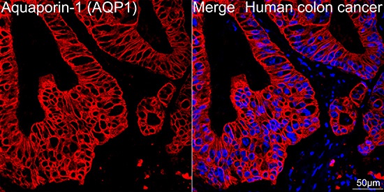
Confocal imaging of paraffin-embedded Human colon cancer tissue using Aquaporin-1 (AQP1) Rabbit mAb (CAB4195, dilution 1:100) followed by a further incubation with Cy3 Goat Anti-Rabbit IgG (H+L) (CABS007, dilution 1:500) (Red). DAPI was used for nuclear staining (Blue). High pressure antigen retrieval performed with 0.01M Citrate Buffer (pH 6.0) prior to IF staining. Objective: 40x.
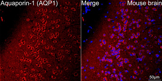
Confocal imaging of paraffin-embedded Mouse brain tissue using Aquaporin-1 (AQP1) Rabbit mAb (CAB4195, dilution 1:100) followed by a further incubation with Cy3 Goat Anti-Rabbit IgG (H+L) (CABS007, dilution 1:500) (Red). DAPI was used for nuclear staining (Blue). Microwave antigen retrieval performed with 0.01M Citrate Buffer (pH 6.0) prior to IF staining. Objective: 40x.


")
")

