The Arp3 Monoclonal Antibody (CAB4514) is a high-quality antibody developed for reliable detection and analysis of target proteins. This highly specific antibody, developed using rabbit monoclonal technology, provides reliable and reproducible results in a variety of applications, including Western blot, immunofluorescence, and immunoprecipitation.ARP3 is essential for cellular processes such as cell motility, cell shape changes, and intracellular transport, making it a crucial target for investigation in cell biology and cancer research.
This antibody is validated for use in WB, IHC-P, IF/ICC, ELISA applications and has demonstrated reactivity against Human, Mouse, Rat samples.
Product Name:
Arp3 Monoclonal Antibody
SKU:
CAB4514
Size:
20μL, 100μL
Reactivity:
Human, Mouse, Rat
Clone Number:
ARC1067
Conjugate:
Unconjugated
Immunogen:
Synthetic peptide. This information is considered to be commercially sensitive.
Recommended starting concentration is 1 μg/mL. Please optimize the concentration based on your specific assay requirements
Synonyms:
ARP3, Arp3
Positive Sample:
HepG2, Jurkat, U-251MG, Mouse lung, Mouse liver, Mouse spleen, Rat lung, Rat brain, Rat spleen
Cellular Localization:
Cell Projection, Cytoplasm, Cytoskeleton.
Calculated MW:
47kDa
Observed MW:
47kDa
The specific function of this gene has not yet been determined; however, the protein it encodes is known to be a major constituent of the ARP2/3 complex. This complex is located at the cell surface and is essential to cell shape and motility through lamellipodial actin assembly and protrusion. Three transcript variants encoding two different isoforms have been found for this gene.
Purification Method
Affinity purification
Gene ID
10096
RRID
AB_2863284
Buffer Information
Store at -20℃. Avoid freeze / thaw cycles. Buffer: PBS containing 50% glycerol and 0.05% BSA, preserved with proclin300 or sodium azide, pH 7.3.
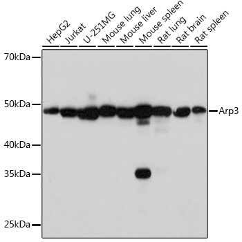
Western blot analysis of various lysates using Arp3 Rabbit mAb (CAB4514) at 1:1000 dilution. Secondary antibody: HRP-conjugated Goat anti-Rabbit IgG (H+L) (CABS014) at 1:10000 dilution. Lysates/proteins: 25μg per lane. Blocking buffer: 3% nonfat dry milk in TBST. Detection: ECL Basic Kit (AbGn00020). Exposure time: 3s.
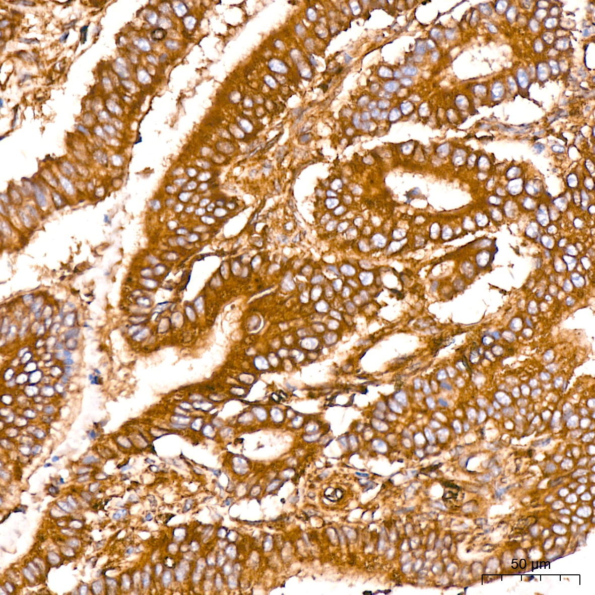
Immunohistochemistry analysis of paraffin-embedded Human colon carcinoma tissue using Arp3 Rabbit mAb (CAB4514) at a dilution of 1:200 (40x lens). High pressure antigen retrieval performed with 0.01M Citrate buffer (pH 6.0) prior to IHC staining.
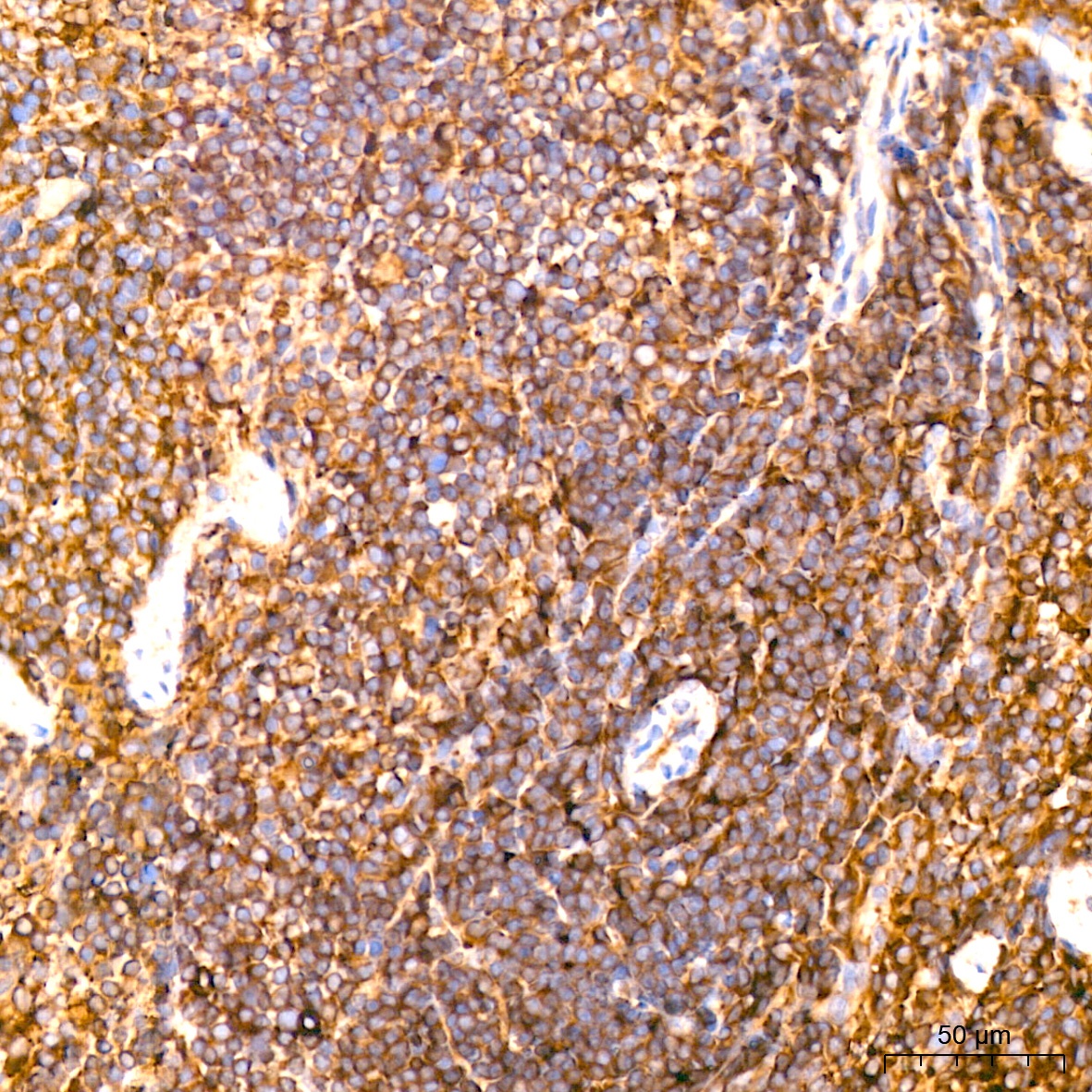
Immunohistochemistry analysis of paraffin-embedded Mouse spleen tissue using Arp3 Rabbit mAb (CAB4514) at a dilution of 1:200 (40x lens). High pressure antigen retrieval performed with 0.01M Citrate buffer (pH 6.0) prior to IHC staining.
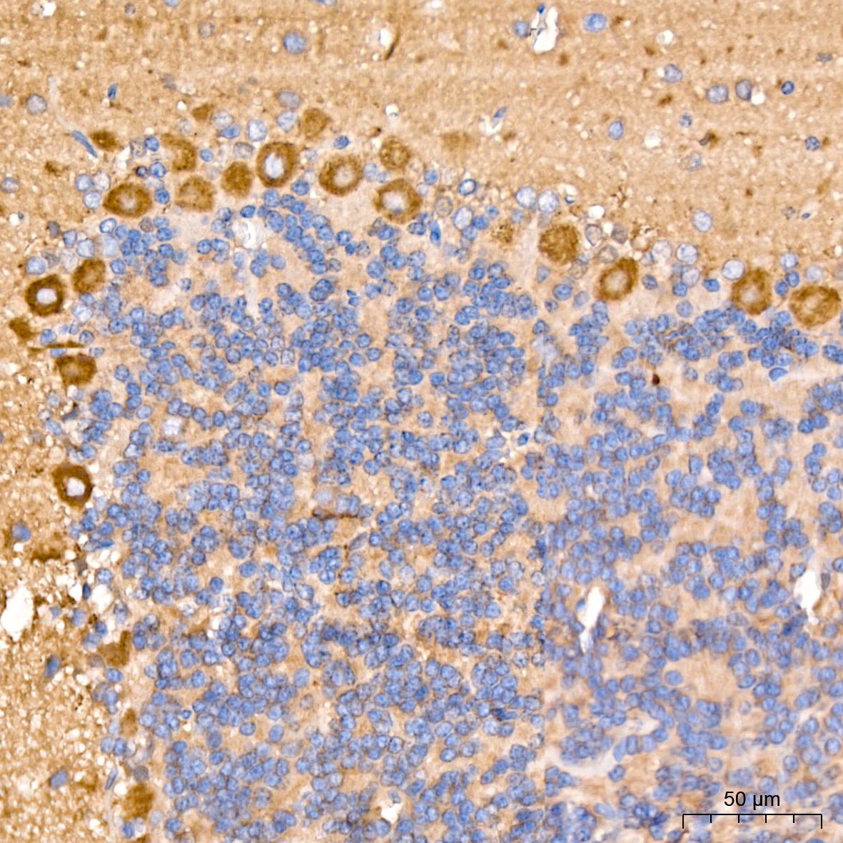
Immunohistochemistry analysis of paraffin-embedded Rat brain tissue using Arp3 Rabbit mAb (CAB4514) at a dilution of 1:200 (40x lens). High pressure antigen retrieval performed with 0.01M Citrate buffer (pH 6.0) prior to IHC staining.
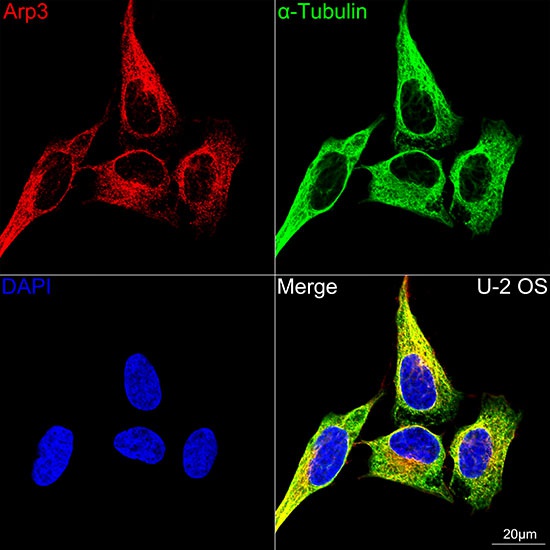
Confocal imaging of U-2 OS cells using Arp3 Rabbit mAb (CAB4514,dilution 1:100)(Red). The cells were counterstained with α-Tubulin Mouse mAb (AC012,dilution 1:400) (Green). DAPI was used for nuclear staining (blue). Objective: 100x.