The ART1 Antibody (CAB10103) is a high-quality antibody developed for reliable detection and analysis of target proteins. This antibody, produced in rabbits, demonstrates high reactivity with human samples and is validated for Western blot applications. By specifically binding to the ART1 protein, researchers can easily detect and analyze its expression in a variety of cell types, making it ideal for studies in immunology, metabolism, and cancer research.ART1, also known as ADP-ribosyltransferase 1, is involved in the post-translational modification of proteins by adding ADP-ribose moieties. This process plays a crucial role in various cellular functions, including immune response regulation and signal transduction pathways.
This antibody is validated for use in WB, IHC-P, IF/ICC, ELISA applications and has demonstrated reactivity against Human, Mouse samples.
Product Name:
ART1 Antibody
SKU:
CAB10103
Size:
20μL, 100μL
Reactivity:
Human, Mouse
Conjugate:
Unconjugated
Immunogen:
Recombinant protein (or fragment).This information is considered to be commercially sensitive.
ADP-ribosyltransferase catalyzes the ADP-ribosylation of arginine residues in proteins. Mono-ADP-ribosylation is a posttranslational modification of proteins that is interfered with by a variety of bacterial toxins including cholera, pertussis, and heat-labile enterotoxins of E. coli. The amino acid sequence consists of predominantly hydrophobic N- and C-terminal regions, which is characteristic of glycosylphosphatidylinositol (GPI)-anchored proteins. This gene was previously designated ART2.
Purification Method
Affinity purification
Gene ID
417
RRID
AB_2757626
Buffer Information
Store at -20℃. Avoid freeze / thaw cycles. Buffer: PBS containing 50% glycerol, preserved with proclin300 or sodium azide, pH 7.3.
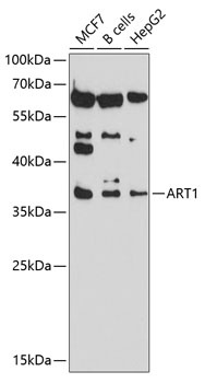
Western blot analysis of various lysates using ART1 Rabbit pAb (CAB10103) at 1:1000 dilution. Secondary antibody: HRP-conjugated Goat anti-Rabbit IgG (H+L) (CABS014) at 1:10000 dilution. Lysates/proteins: 25μg per lane. Blocking buffer: 3% nonfat dry milk in TBST. Detection: ECL Basic Kit (AbGn00020). Exposure time: 90s.
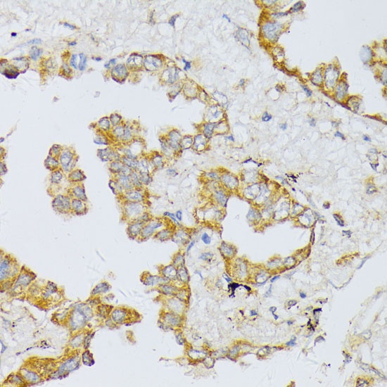
Immunohistochemistry analysis of paraffin-embedded Human lung cancer using ART1 Rabbit pAb (CAB10103) at dilution of 1:100 (40x lens). Microwave antigen retrieval performed with 0.01M PBS Buffer (pH 7.2) prior to IHC staining.
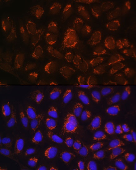
Immunofluorescence analysis of U-2 OS cells using ART1 Rabbit pAb (CAB10103) at dilution of 1:100. Secondary antibody: Cy3-conjugated Goat anti-Rabbit IgG (H+L) (CABS007) at 1:500 dilution. Blue: DAPI for nuclear staining.