The ASCC2 Antibody (CAB13789) is a high-quality antibody developed for reliable detection and analysis of target proteins. This antibody, produced in rabbits, has high reactivity with human samples and is validated for use in Western blot applications. By binding to ASCC2 protein, this antibody allows for accurate detection and analysis in various cell types, making it an essential reagent for studies in cancer biology and genomic stability.ASCC2 is known to play a crucial role in the DNA damage response pathway, contributing to the repair of DNA lesions and ensuring the integrity of the genome.
This antibody is validated for use in WB, ELISA applications and has demonstrated reactivity against Human, Mouse, Rat samples.
Product Name:
ASCC2 Antibody
SKU:
CAB13789
Size:
20μL, 100μL
Reactivity:
Human, Mouse, Rat
Conjugate:
Unconjugated
Immunogen:
Recombinant protein (or fragment).This information is considered to be commercially sensitive.
Recommended starting concentration is 1 μg/mL. Please optimize the concentration based on your specific assay requirements.
Synonyms:
p100, ASC1p100, ASCC2
Positive Sample:
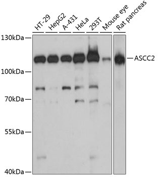
HT-29, HepG2, A-431, HeLa, 293T, Mouse eye, Rat pancreas
Cellular Localization:
Nuclear Speck, Nucleoplasm, Nucleus.
Calculated MW:
86kDa
Observed MW:
110kDa
Predicted to enable ubiquitin binding activity. Involved in regulation of transcription, DNA-templated; rescue of stalled ribosome; and ribosome-associated ubiquitin-dependent protein catabolic process. Located in nucleus. Part of activating signal cointegrator 1 complex.
Purification Method
Affinity purification
Gene ID
84164
RRID
AB_2760647
Buffer Information
Store at -20℃. Avoid freeze / thaw cycles. Buffer: PBS with 0.01% thimerosal,50% glycerol,pH7.3.
Western blot analysis of various lysates using ASCC2 Rabbit pAb (CAB13789) at 1:3000 dilution. Secondary antibody: HRP-conjugated Goat anti-Rabbit IgG (H+L) (CABS014) at 1:10000 dilution. Lysates/proteins: 25μg per lane. Blocking buffer: 3% nonfat dry milk in TBST. Detection: ECL Basic Kit (AbGn00020). Exposure time: 60s.