The ATAD3A Antibody (CAB8230) is a high-quality antibody developed for reliable detection and analysis of target proteins. This polyclonal antibody, generated in rabbits, offers high specificity and sensitivity for detecting ATAD3A in human samples, making it suitable for various applications including Western blotting.ATAD3A is known for its involvement in mitochondrial nucleoid organization and homeostasis, as well as its interactions with other mitochondrial proteins. Dysfunction of ATAD3A has been linked to mitochondrial disorders and metabolic diseases, highlighting the importance of studying this protein in the context of human health and disease.
This antibody is validated for use in WB, IHC-P, ELISA applications and has demonstrated reactivity against Human, Mouse, Rat samples.
Product Name:
ATAD3A Antibody
SKU:
CAB8230
Size:
20μL, 100μL
Reactivity:
Human, Mouse, Rat
Conjugate:
Unconjugated
Immunogen:
Recombinant protein (or fragment).This information is considered to be commercially sensitive.
This gene encodes a ubiquitously expressed mitochondrial membrane protein that contributes to mitochondrial dynamics, nucleoid organization, protein translation, cell growth, and cholesterol metabolism. This gene is a member of the ATPase family AAA-domain containing 3 gene family which, in humans, includes two other paralogs. Naturally occurring mutations in this gene are associated with distinct neurological syndromes including Harel-Yoon syndrome. High-level expression of this gene is associated with poor survival in breast cancer patients. A homozygous knockout of the orthologous gene in mice results in embryonic lethality at day 7.5 due to growth retardation and defective development of the trophoblast lineage. Alternative splicing results in multiple transcript variants.
Purification Method
Affinity purification
Gene ID
55210
RRID
AB_2768483
Buffer Information
Store at -20℃. Avoid freeze / thaw cycles. Buffer: PBS containing 50% glycerol, preserved with proclin300 or sodium azide, pH 7.3.
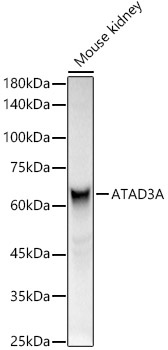
Western blot analysis of lysates from Mouse kidney, using ATAD3A Rabbit pAb (CAB8230) at 1:400 dilution. Secondary antibody: HRP-conjugated Goat anti-Rabbit IgG (H+L) (CABS014) at 1:10000 dilution. Lysates/proteins: 25μg per lane. Blocking buffer: 3% nonfat dry milk in TBST. Detection: ECL Basic Kit (AbGn00020). Exposure time: 3s.
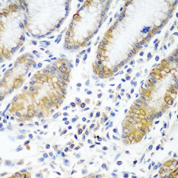
Immunohistochemistry analysis of paraffin-embedded Human stomach using ATAD3A Rabbit pAb (CAB8230) at dilution of 1:100 (40x lens). Microwave antigen retrieval performed with 0.01M PBS Buffer (pH 7.2) prior to IHC staining.