The Ataxin 3 Antibody (CAB1243) is a high-quality antibody developed for reliable detection and analysis of target proteins. This antibody, produced in rabbits, has high specificity for human samples and is suitable for use in Western blotting and immunohistochemistry applications.Ataxin-3 is a key player in the pathogenesis of SCA3, a progressive disorder affecting coordination and movement control. By targeting Ataxin-3, researchers can investigate the mechanisms underlying disease development and progression, potentially leading to new therapeutic strategies for managing SCA3 and other related conditions.
This antibody is validated for use in WB, IHC-P, IF/ICC, ELISA applications and has demonstrated reactivity against Human, Mouse, Rat samples.
Product Name:
Ataxin 3 Antibody
SKU:
CAB1243
Size:
20μL, 100μL
Reactivity:
Human, Mouse, Rat
Conjugate:
Unconjugated
Immunogen:
Recombinant protein (or fragment).This information is considered to be commercially sensitive.
Recommended starting concentration is 1 μg/mL. Please optimize the concentration based on your specific assay requirements.
Synonyms:
AT3, JOS, MJD, ATX3, MJD1, SCA3, Ataxin-3 (ATXN3)
Positive Sample:
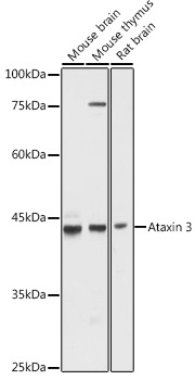
Mouse brain, Mouse thymus, Rat brain
Cellular Localization:
Nucleus Matrix.
Calculated MW:
41kDa
Observed MW:
42kDa
Machado-Joseph disease, also known as spinocerebellar ataxia-3, is an autosomal dominant neurologic disorder. The protein encoded by this gene contains (CAG)n repeats in the coding region, and the expansion of these repeats from the normal 12-44 to 52-86 is one cause of Machado-Joseph disease. There is a negative correlation between the age of onset and CAG repeat numbers. Alternatively spliced transcript variants encoding different isoforms have been described for this gene.
Purification Method
Affinity purification
Gene ID
4287
RRID
AB_2759271
Buffer Information
Store at -20℃. Avoid freeze / thaw cycles. Buffer: PBS containing 50% glycerol, preserved with proclin300 or sodium azide, pH 7.3.
Western blot analysis of various lysates using Ataxin-3 (ATXN3) Rabbit pAb (CAB1243) at 1:1000 dilution. Secondary antibody: HRP-conjugated Goat anti-Rabbit IgG (H+L) (CABS014) at 1:10000 dilution. Lysates/proteins: 25μg per lane. Blocking buffer: 3% nonfat dry milk in TBST. Detection: ECL Basic Kit (AbGn00020). Exposure time: 10s.
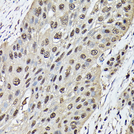
Immunohistochemistry analysis of paraffin-embedded Human esophageal cancer using Ataxin-3 (ATXN3) Rabbit pAb (CAB1243) at dilution of 1:100 (40x lens). High pressure antigen retrieval performed with 0.01M Citrate buffer (pH 6.0) prior to IHC staining.
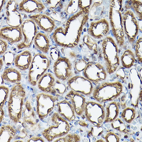
Immunohistochemistry analysis of paraffin-embedded Rat kidney using Ataxin-3 (ATXN3) Rabbit pAb (CAB1243) at dilution of 1:100 (40x lens). High pressure antigen retrieval performed with 0.01M Citrate buffer (pH 6.0) prior to IHC staining.
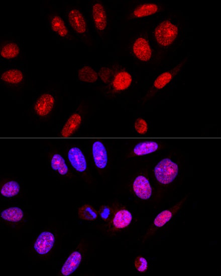
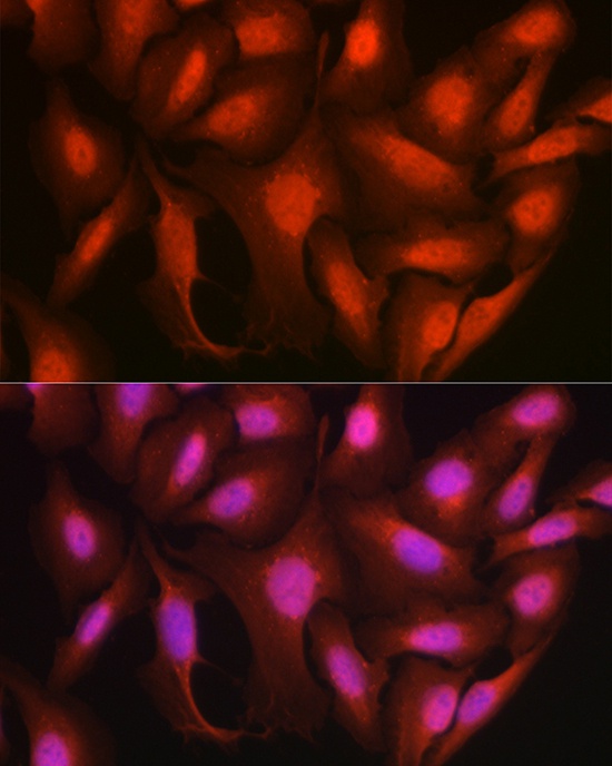
Confocal immunofluorescence analysis of U-2 OS cells using Ataxin-3 (ATXN3) Rabbit pAb (CAB1243) at dilution of 1:100. Blue: DAPI for nuclear staining.
Immunofluorescence analysis of U2OS cells using Ataxin-3 (ATXN3) Rabbit pAb (CAB1243) at dilution of 1:100 (40x lens). Secondary antibody: Cy3-conjugated Goat anti-Rabbit IgG (H+L) (CABS007) at 1:500 dilution. Blue: DAPI for nuclear staining.