The ATG10 Monoclonal Antibody (CAB6848) is a high-quality antibody developed for reliable detection and analysis of target proteins. This antibody, raised in rabbits, is validated for use in various applications such as Western blot, immunohistochemistry, and immunofluorescence, allowing for the detection and analysis of ATG10 in different cell types and tissues.ATG10 is a critical component of the autophagy process, responsible for the conjugation of ATG12 to ATG5, which is essential for the formation of the autophagosome. Dysregulation of autophagy has been linked to various diseases including cancer, neurodegenerative disorders, and metabolic conditions.
This antibody is validated for use in WB, IF/ICC, ELISA applications and has demonstrated reactivity against Human, Mouse samples.
Product Name:
ATG10 Monoclonal Antibody
SKU:
CAB6848
Size:
20μL, 100μL
Reactivity:
Human, Mouse
Clone Number:
ARC1425
Conjugate:
Unconjugated
Immunogen:
Synthetic peptide. This information is considered to be commercially sensitive.
Recommended starting concentration is 1 μg/mL. Please optimize the concentration based on your specific assay requirements.
Synonyms:
APG10, APG10L, pp12616, ATG10
Positive Sample:
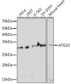
HeLa, 293T, K-562, SH-SY5Y, Mouse heart
Cellular Localization:
Cytoplasm.
Calculated MW:
25kDa
Observed MW:
25kDa
Autophagy is a process for the bulk degradation of cytosolic compartments by lysosomes. ATG10 is an E2-like enzyme involved in 2 ubiquitin-like modifications essential for autophagosome formation: ATG12 (MIM 609608)-ATG5 (MIM 604261) conjugation and modification of a soluble form of MAP-LC3 (MAP1LC3A; MIM 601242), a homolog of yeast Apg8, to a membrane-bound form (Nemoto et al., 2003 [PubMed 12890687]).
Purification Method
Affinity purification
Gene ID
83734
RRID
AB_2863543
Buffer Information
Store at -20℃. Avoid freeze / thaw cycles. Buffer: PBS containing 50% glycerol and 0.05% BSA, preserved with proclin300 or sodium azide, pH 7.3.
Western blot analysis of various lysates using ATG10 Rabbit mAb (CAB6848) at 1:1000 dilution. Secondary antibody: HRP-conjugated Goat anti-Rabbit IgG (H+L) (CABS014) at 1:10000 dilution. Lysates/proteins: 25μg per lane. Blocking buffer: 3% nonfat dry milk in TBST. Detection: ECL Basic Kit (AbGn00020). Exposure time: 3min.
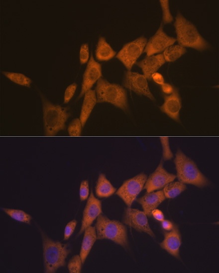
Immunofluorescence analysis of NIH-3T3 cells using ATG10 Rabbit mAb (CAB6848) at dilution of 1:100 (40x lens). Secondary antibody: Cy3-conjugated Goat anti-Rabbit IgG (H+L) (CABS007) at 1:500 dilution. Blue: DAPI for nuclear staining.