The ATG10 Monoclonal Antibody (CAB6848) is a high-quality antibody developed for reliable detection and analysis of target proteins. Autophagy is a process for the bulk degradation of cytosolic compartments by lysosomes. ATG10 is an E2-like enzyme involved in 2 ubiquitin-like modifications essential for autophagosome formation: ATG12 (MIM 609608)-ATG5 (MIM 604261) conjugation and modification of a soluble form of MAP-LC3 (MAP1LC3A; MIM 601242), a homolog of yeast Apg8, to a membrane-bound form (Nemoto et al., 2003 [PubMed 12890687]).
This antibody is validated for use in WB, IF/ICC, ELISA applications and has demonstrated reactivity against Human, Mouse samples.
Product Name:
ATG10 Monoclonal Antibody
SKU:
CAB6848
Size:
100μL, 20μL
Reactivity:
Human, Mouse
Clone Number:
ARC1425
Conjugate:
Unconjugated
Immunogen:
Synthetic peptide. This information is considered to be commercially sensitive.
Tested Applications:
WBIF/ICCELISA
Recommended Dilution:
WB
1:500 - 1:2000
IF/ICC
1:50 - 1:200
ELISA
Recommended starting concentration is 1 μg/mL. Please optimize the concentration based on your specific assay requirements.
Synonyms:
APG10, APG10L, pp12616, ATG10
Positive Sample:
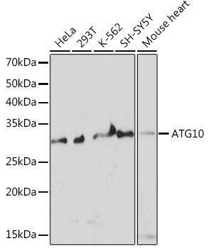
HeLa, 293T, K-562, SH-SY5Y, Mouse heart
Cellular Localization:
Cytoplasm.
Calculated MW:
25kDa
Observed MW:
25kDa
Autophagy is a process for the bulk degradation of cytosolic compartments by lysosomes. ATG10 is an E2-like enzyme involved in 2 ubiquitin-like modifications essential for autophagosome formation: ATG12 (MIM 609608)-ATG5 (MIM 604261) conjugation and modification of a soluble form of MAP-LC3 (MAP1LC3A; MIM 601242), a homolog of yeast Apg8, to a membrane-bound form (Nemoto et al., 2003 [PubMed 12890687]).
Purification Method
Affinity purification
Gene ID
83734
RRID
AB_2863543
Buffer Information
Store at -20℃. Avoid freeze / thaw cycles. Buffer: PBS containing 50% glycerol and 0.05% BSA, preserved with proclin300 or sodium azide, pH 7.3.
Western blot analysis of various lysates using ATG10 Rabbit mAb (CAB6848) at 1:1000 dilution. Secondary antibody: HRP-conjugated Goat anti-Rabbit IgG (H+L) (AS014) at 1:10000 dilution. Lysates/proteins: 25μg per lane. Blocking buffer: 3% nonfat dry milk in TBST. Detection: ECL Basic Kit (AbGn00020). Exposure time: 3min.
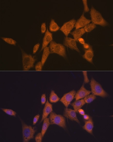
Immunofluorescence analysis of NIH-3T3 cells using ATG10 Rabbit mAb (CAB6848) at dilution of 1:100 (40x lens). Secondary antibody: Cy3-conjugated Goat anti-Rabbit IgG (H+L) (AS007) at 1:500 dilution. Blue: DAPI for nuclear staining.