The ATG12 Antibody (CAB15609) is a high-quality antibody developed for reliable detection and analysis of target proteins. This antibody, raised in rabbits, demonstrates high reactivity with human samples and has been validated for use in Western blot applications. By specifically binding to the ATG12 protein, this antibody enables accurate detection and analysis in a variety of cell types, making it ideal for studying autophagy and its role in various physiological and pathological processes.ATG12 is a key component of the autophagy pathway, a cellular process crucial for maintaining cellular homeostasis by degrading damaged organelles and proteins.
This antibody is validated for use in WB, IF/ICC, ELISA applications and has demonstrated reactivity against Human, Mouse, Rat samples.
Product Name:
ATG12 Antibody
SKU:
CAB15609
Size:
20μL, 100μL
Reactivity:
Human, Mouse, Rat
Conjugate:
Unconjugated
Immunogen:
Synthetic peptide. This information is considered to be commercially sensitive.
Autophagy is a process of bulk protein degradation in which cytoplasmic components, including organelles, are enclosed in double-membrane structures called autophagosomes and delivered to lysosomes or vacuoles for degradation. ATG12 is the human homolog of a yeast protein involved in autophagy (Mizushima et al., 1998 [PubMed 9852036]).
Purification Method
Affinity purification
Gene ID
9140
RRID
AB_2763015
Buffer Information
Store at -20℃. Avoid freeze / thaw cycles. Buffer: PBS containing 50% glycerol, preserved with proclin300 or sodium azide, pH 7.3.
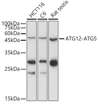
Western blot analysis of various lysates using ATG12 Rabbit pAb (CAB15609) at 1:1000 dilution. Secondary antibody: HRP-conjugated Goat anti-Rabbit IgG (H+L) (CABS014) at 1:10000 dilution. Lysates/proteins: 25μg per lane. Blocking buffer: 3% nonfat dry milk in TBST. Detection: ECL Enhanced Kit (AbGn00021). Exposure time: 180s.
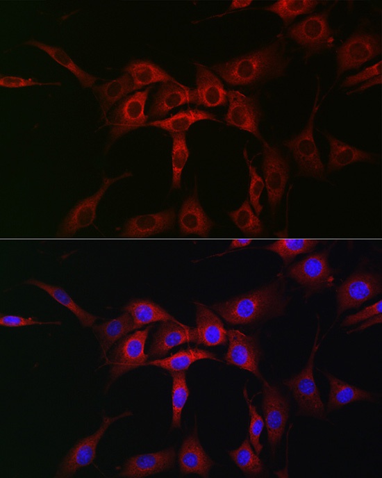
Immunofluorescence analysis of NIH/3T3 cells using ATG12 Rabbit pAb (CAB15609) at dilution of 1:50 (40x lens). Secondary antibody: Cy3-conjugated Goat anti-Rabbit IgG (H+L) (CABS007) at 1:500 dilution. Blue: DAPI for nuclear staining.
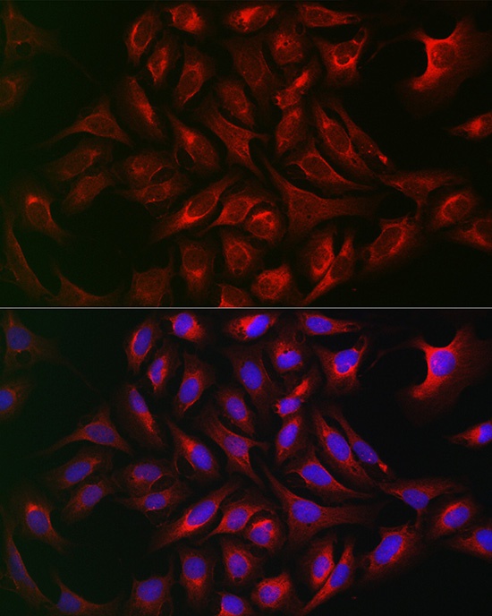
Immunofluorescence analysis of U2OS cells using ATG12 Rabbit pAb (CAB15609) at dilution of 1:50 (40x lens). Secondary antibody: Cy3-conjugated Goat anti-Rabbit IgG (H+L) (CABS007) at 1:500 dilution. Blue: DAPI for nuclear staining.