The ATG16L1 Antibody (CAB11969) is a high-quality antibody developed for reliable detection and analysis of target proteins. The protein encoded by this gene is part of a large protein complex that is necessary for autophagy, the major process by which intracellular components are targeted to lysosomes for degradation. Defects in this gene are a cause of susceptibility to inflammatory bowel disease type 10 (IBD10). Several transcript variants encoding different isoforms have been found for this gene. RRID AB_2758902 Gene ID 55054 Swiss Prot Synonym IBD10; WDR30; APG16L; ATG16A; ATG16L; ATG16L1
This antibody is validated for use in WB, IHC-P, IF/ICC, ELISA applications and has demonstrated reactivity against Human, Mouse, Rat samples.
Product Name:
ATG16L1 Antibody
SKU:
CAB11969
Size:
100μL, 20μL
Reactivity:
Human, Mouse, Rat
Clone Number:
-
Conjugate:
Unconjugated
Immunogen:
Synthetic peptide. This information is considered to be commercially sensitive.
Tested Applications:
WBIHC-PIF/ICCELISA
Recommended Dilution:
WB
1:500 - 1:2000
IHC-P
1:50 - 1:200
IF
/
ICC
1:50 - 1:200
ELISA
Recommended starting concentration is 1 μg/mL. Please optimize the concentration based on your specific assay requirements.
The protein encoded by this gene is part of a large protein complex that is necessary for autophagy, the major process by which intracellular components are targeted to lysosomes for degradation. Defects in this gene are a cause of susceptibility to inflammatory bowel disease type 10 (IBD10). Several transcript variants encoding different isoforms have been found for this gene. RRID AB_2758902 Gene ID 55054 Swiss Prot Synonym IBD10; WDR30; APG16L; ATG16A; ATG16L; ATG16L1
Purification Method:
Affinity purification
Gene ID:
55054
RRID:
AB_2758902
Buffer Information:
Store at -20℃. Avoid freeze / thaw cycles. Buffer: PBS with 0.01% thimerosal,50% glycerol,pH7.3.
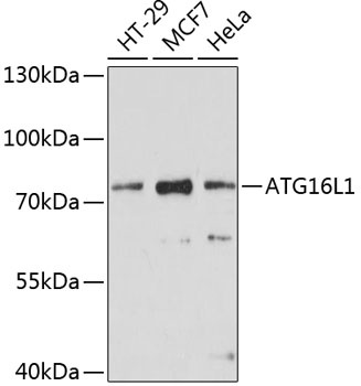
Western blot analysis of various lysates using ATG16L1 Rabbit pAb (CAB11969) at 1:1000 dilution. Secondary antibody: HRP-conjugated Goat anti-Rabbit IgG (H+L) (AS014) at 1:10000 dilution. Lysates/proteins: 25μg per lane. Blocking buffer: 3% nonfat dry milk in TBST. Detection: ECL Enhanced Kit (AbGn00021). Exposure time: 30s.
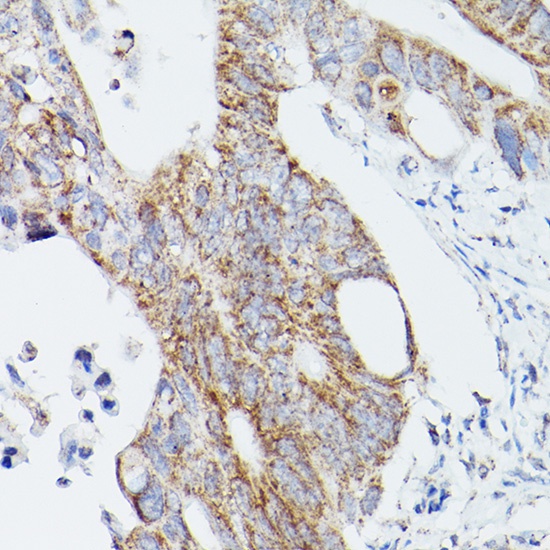
Immunohistochemistry analysis of paraffin-embedded Human colon carcinoma using ATG16L1 Rabbit pAb (CAB11969) at dilution of 1:100 (40x lens). Microwave antigen retrieval performed with 0.01M PBS Buffer (pH 7.2) prior to IHC staining.
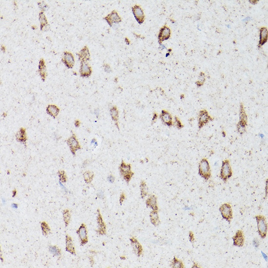
Immunohistochemistry analysis of paraffin-embedded Mouse brain using ATG16L1 Rabbit pAb (CAB11969) at dilution of 1:100 (40x lens). Microwave antigen retrieval performed with 0.01M PBS Buffer (pH 7.2) prior to IHC staining.
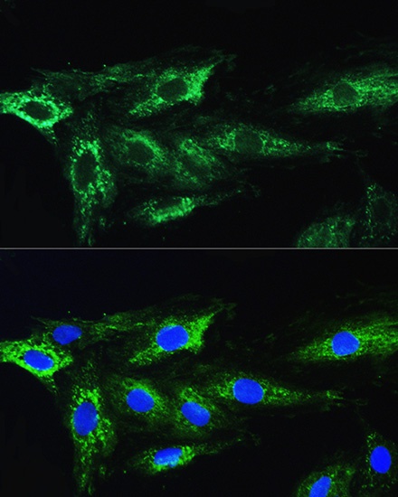
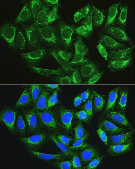
Immunofluorescence analysis of H9C2 cells using ATG16L1 Rabbit pAb (CAB11969) at dilution of 1:100. Blue: DAPI for nuclear staining.
Immunofluorescence analysis of U2OS cells using ATG16L1 Rabbit pAb (CAB11969) at dilution of 1:100. Blue: DAPI for nuclear staining.