The ATG16L1 Antibody (CAB1871) is a high-quality antibody developed for reliable detection and analysis of target proteins. This antibody, generated in rabbits, has been rigorously tested for its reactivity with human samples and is optimized for use in Western blot applications. By targeting the ATG16L1 protein, this antibody allows for the detection and analysis of autophagy-related processes in various cell types, making it essential for studies in cell biology and disease research.
This antibody is validated for use in WB, IHC-P, IF/ICC, ELISA applications and has demonstrated reactivity against Human, Mouse, Rat samples.
Product Name:
ATG16L1 Antibody
SKU:
CAB1871
Size:
20μL, 100μL
Reactivity:
Human, Mouse, Rat
Conjugate:
Unconjugated
Immunogen:
Recombinant protein (or fragment).This information is considered to be commercially sensitive.
The protein encoded by this gene is part of a large protein complex that is necessary for autophagy, the major process by which intracellular components are targeted to lysosomes for degradation. Defects in this gene are a cause of susceptibility to inflammatory bowel disease type 10 (IBD10). Several transcript variants encoding different isoforms have been found for this gene.
Purification Method
Affinity purification
Gene ID
55054
RRID
AB_2763904
Buffer Information
Store at -20℃. Avoid freeze / thaw cycles. Buffer: PBS containing 50% glycerol, preserved with proclin300 or sodium azide, pH 7.3.
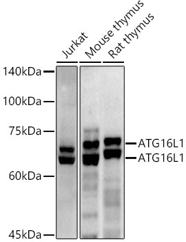
Western blot analysis of various lysates using ATG16L1 Rabbit pAb (CAB1871) at 1:500 dilution. Secondary antibody: HRP-conjugated Goat anti-Rabbit IgG (H+L) (CABS014) at 1:10000 dilution. Lysates/proteins: 25μg per lane. Blocking buffer: 3% nonfat dry milk in TBST. Detection: ECL Enhanced Kit (AbGn00021). Exposure time: 180s.
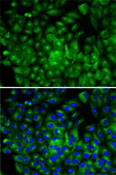
Immunofluorescence analysis of A-549 cells using ATG16L1 Rabbit pAb (CAB1871). Secondary antibody: Cy3-conjugated Goat anti-Rabbit IgG (H+L) (CABS007) at 1:500 dilution. Blue: DAPI for nuclear staining.