The ATG16L1 Monoclonal Antibody (CAB3637) is a high-quality antibody developed for reliable detection and analysis of target proteins. This antibody, generated from rabbit-derived mononclonal cells, exhibits high specificity and reactivity with human samples, making it ideal for use in Western blot applications.ATG16L1 is a crucial component of the autophagy pathway, contributing to the degradation of intracellular components and the maintenance of cellular health. Dysregulation of ATG16L1 has been implicated in various diseases, including inflammatory disorders, infectious diseases, and cancer.
This antibody is validated for use in WB, ELISA applications and has demonstrated reactivity against Human, Mouse samples.
Product Name:
ATG16L1 Monoclonal Antibody
SKU:
CAB3637
Size:
20μL, 100μL
Reactivity:
Human, Mouse
Clone Number:
ARC0812
Conjugate:
Unconjugated
Immunogen:
Recombinant protein (or fragment).This information is considered to be commercially sensitive.
The protein encoded by this gene is part of a large protein complex that is necessary for autophagy, the major process by which intracellular components are targeted to lysosomes for degradation. Defects in this gene are a cause of susceptibility to inflammatory bowel disease type 10 (IBD10). Several transcript variants encoding different isoforms have been found for this gene.
Purification Method
Affinity purification
Gene ID
55054
RRID
AB_2863103
Buffer Information
Store at -20℃. Avoid freeze / thaw cycles. Buffer: PBS containing 50% glycerol and 0.05% BSA, preserved with proclin300 or sodium azide, pH 7.3.
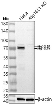
Western blot analysis of lysates from wild type (WT) and ATG16L1 knockout (KO) HeLa cells using [KO Validated] ATG16L1 Rabbit mAb (CAB3637) at 1:1000 dilution incubated overnight at 4℃. Secondary antibody: HRP-conjugated Goat anti-Rabbit IgG (H+L) (CABS014) at 1:10000 dilution. Lysates/proteins: 25 μg per lane. Blocking buffer: 3% nonfat dry milk in TBST. Detection: ECL Basic Kit (AbGn00020). Exposure time: 10s.
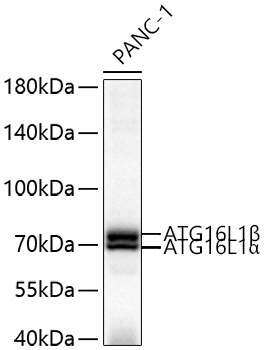
Western blot analysis of lysates from PANC-1 cells using [KO Validated] ATG16L1 Rabbit mAb (CAB3637) at 1:1000 dilution incubated overnight at 4℃. Secondary antibody: HRP-conjugated Goat anti-Rabbit IgG (H+L) (CABS014) at 1:10000 dilution. Lysates/proteins: 25 μg per lane. Blocking buffer: 3% nonfat dry milk in TBST. Detection: ECL Basic Kit (AbGn00020). Exposure time: 20 s.