The ATG3 Antibody (CAB5809) is a high-quality antibody developed for reliable detection and analysis of target proteins. This antibody, generated in rabbits, exhibits high specificity and sensitivity towards human samples, making it an ideal choice for Western blot applications.ATG3 is a key player in the autophagy pathway, responsible for facilitating the conjugation of ATG8 to phosphatidylethanolamine on the autophagosome membrane. By targeting ATG3 with this polyclonal antibody, researchers can visualize and quantify ATG3 protein levels in various cell types, aiding in the investigation of autophagy-related processes.
This antibody is validated for use in WB, IF/ICC, ELISA applications and has demonstrated reactivity against Human, Mouse, Rat samples.
Product Name:
ATG3 Antibody
SKU:
CAB5809
Size:
20μL, 100μL
Reactivity:
Human, Mouse, Rat
Conjugate:
Unconjugated
Immunogen:
Recombinant protein (or fragment).This information is considered to be commercially sensitive.
Recommended starting concentration is 1 μg/mL. Please optimize the concentration based on your specific assay requirements.
Synonyms:
APG3, APG3L, hApg3, PC3-96, APG3-LIKE, ATG3
Positive Sample:
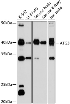
K-562, U-87MG, Mouse brain, Mouse kidney, Rat testis
Cellular Localization:
Cytoplasm.
Calculated MW:
36kDa
Observed MW:
40kDa
This gene encodes a ubiquitin-like-conjugating enzyme and is a component of ubiquitination-like systems involved in autophagy, the process of degradation, turnover and recycling of cytoplasmic constituents in eukaryotic cells. This protein is known to play a role in regulation of autophagy during cell death. A pseudogene of this gene is located on chromosome 20. Alternative splicing results in multiple transcript variants encoding different isoforms.
Purification Method
Affinity purification
Gene ID
64422
RRID
AB_2766561
Buffer Information
Store at -20℃. Avoid freeze / thaw cycles. Buffer: PBS containing 50% glycerol, preserved with proclin300 or sodium azide, pH 7.3.
Western blot analysis of various lysates using ATG3 Rabbit pAb (CAB5809) at 1:1000 dilution. Secondary antibody: HRP-conjugated Goat anti-Rabbit IgG (H+L) (CABS014) at 1:10000 dilution. Lysates/proteins: 25μg per lane. Blocking buffer: 3% nonfat dry milk in TBST. Detection: ECL Basic Kit (AbGn00020). Exposure time: 180s.
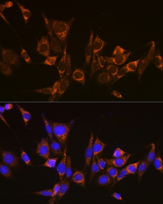
Immunofluorescence analysis of NIH-3T3 cells using ATG3 Rabbit pAb (CAB5809) at dilution of 1:100 (40x lens). Secondary antibody: Cy3-conjugated Goat anti-Rabbit IgG (H+L) (CABS007) at 1:500 dilution. Blue: DAPI for nuclear staining.