The ATG3 Monoclonal Antibody (CAB19594) is a high-quality antibody developed for reliable detection and analysis of target proteins. This highly specific antibody, generated from rabbit immunizations, is optimized for detecting and analyzing ATG3 protein levels in a variety of samples, but is particularly effective in human samples.ATG3 is a crucial component of the autophagy pathway, responsible for conjugating ATG8-like proteins to phosphatidylethanolamine, facilitating the formation of autophagosomes. Dysregulation of autophagy has been implicated in various diseases, including neurodegenerative disorders, cancer, and infectious diseases.
This antibody is validated for use in WB, IHC-P, ELISA applications and has demonstrated reactivity against Human, Mouse, Rat samples.
Product Name:
ATG3 Monoclonal Antibody
SKU:
CAB19594
Size:
20μL, 100μL
Reactivity:
Human, Mouse, Rat
Clone Number:
ARC0073
Conjugate:
Unconjugated
Immunogen:
Synthetic peptide. This information is considered to be commercially sensitive.
Recommended starting concentration is 1 μg/mL. Please optimize the concentration based on your specific assay requirements.
Synonyms:
APG3, APG3L, hApg3, PC3-96, APG3-LIKE, ATG3
Positive Sample:
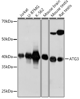
Jurkat, U-87 MG, K-562, Mouse brain, Mouse testis, Rat testis
Cellular Localization:
Cytoplasm.
Calculated MW:
40kDa
Observed MW:
40kDa
This gene encodes a ubiquitin-like-conjugating enzyme and is a component of ubiquitination-like systems involved in autophagy, the process of degradation, turnover and recycling of cytoplasmic constituents in eukaryotic cells. This protein is known to play a role in regulation of autophagy during cell death. A pseudogene of this gene is located on chromosome 20. Alternative splicing results in multiple transcript variants encoding different isoforms.
Purification Method
Affinity purification
Gene ID
64422
RRID
AB_2862687
Buffer Information
Store at -20℃. Avoid freeze / thaw cycles. Buffer: PBS containing 50% glycerol and 0.05% BSA, preserved with proclin300 or sodium azide, pH 7.3.
Western blot analysis of various lysates using ATG3 Rabbit mAb (CAB19594) at 1:1000 dilution. Secondary antibody: HRP-conjugated Goat anti-Rabbit IgG (H+L) (CABS014) at 1:10000 dilution. Lysates/proteins: 25μg per lane. Blocking buffer: 3% nonfat dry milk in TBST. Detection: ECL Basic Kit (AbGn00020). Exposure time: 40s.
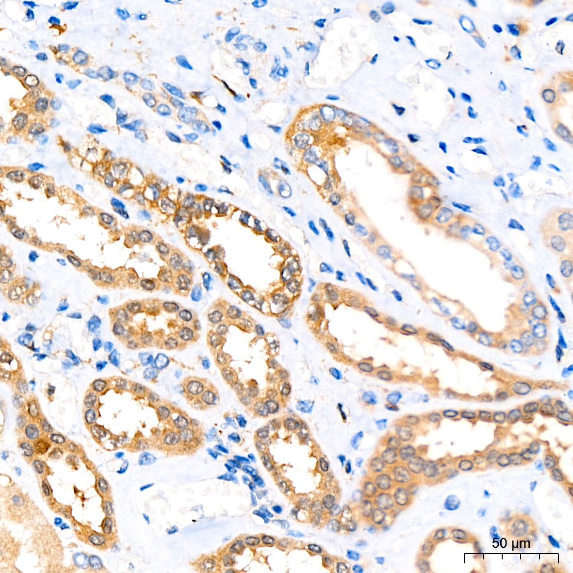
Immunohistochemistry analysis of paraffin-embedded Human kidney tissue using ATG3 Rabbit mAb (CAB19594) at a dilution of 1:200 (40x lens). High pressure antigen retrieval performed with 0.01M Citrate buffer (pH 6.0) prior to IHC staining.
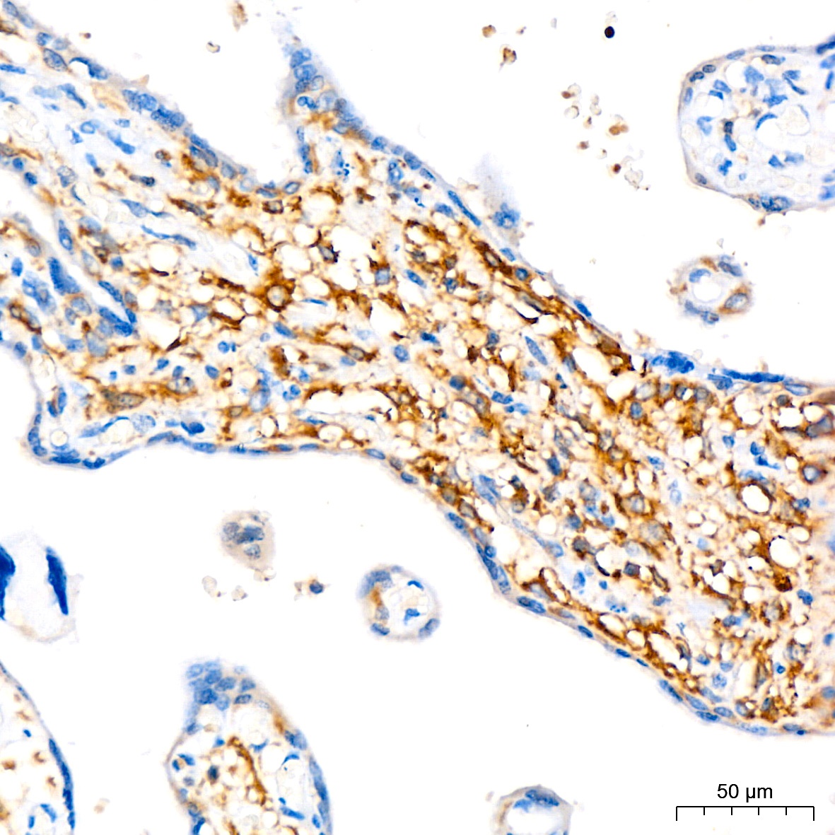
Immunohistochemistry analysis of paraffin-embedded Human placenta tissue using ATG3 Rabbit mAb (CAB19594) at a dilution of 1:200 (40x lens). High pressure antigen retrieval performed with 0.01M Citrate buffer (pH 6.0) prior to IHC staining.
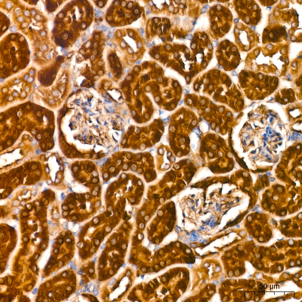
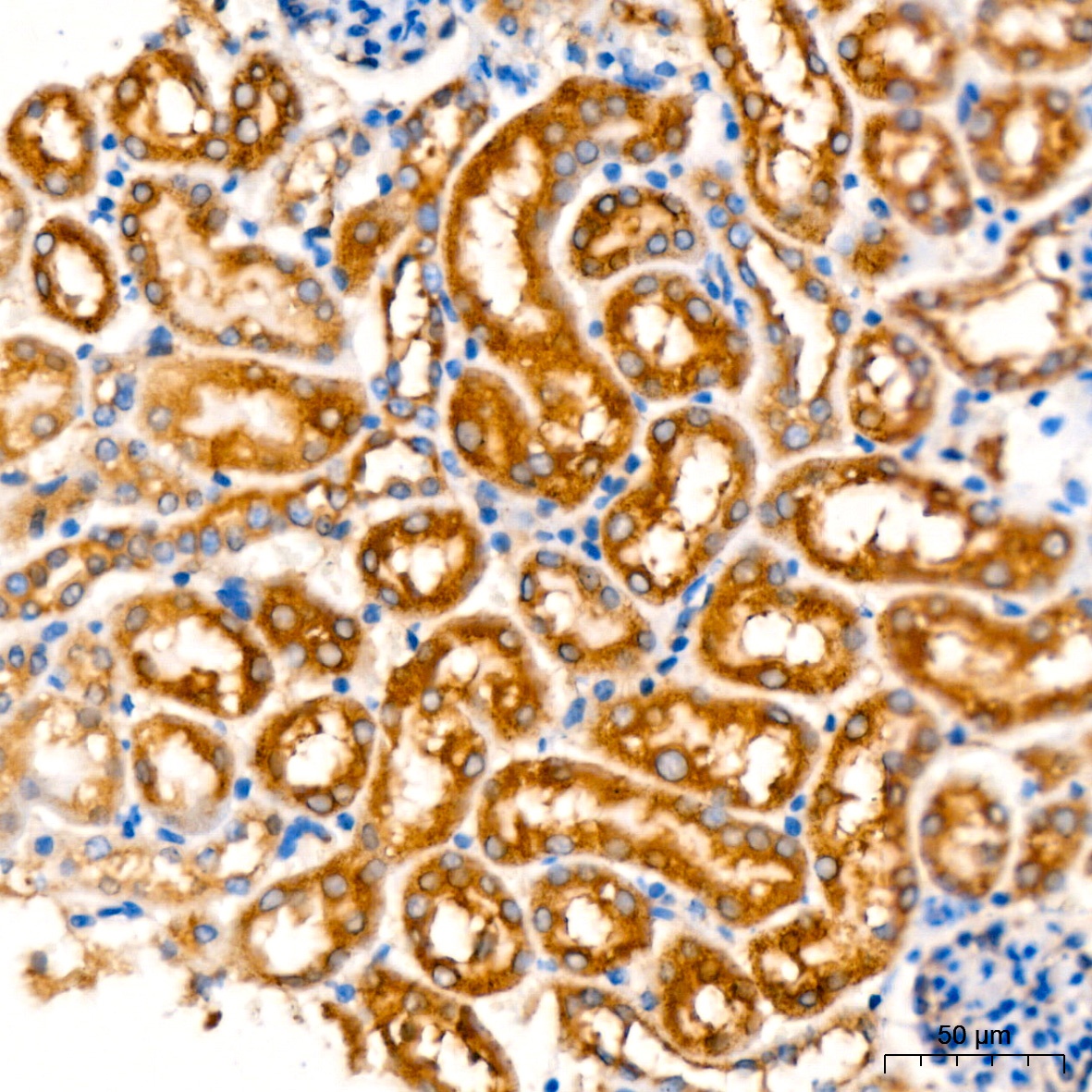
Immunohistochemistry analysis of paraffin-embedded Mouse kidney tissue using ATG3 Rabbit mAb (CAB19594) at a dilution of 1:200 (40x lens). High pressure antigen retrieval performed with 0.01M Tris-EDTA Buffer (pH 9.0) prior to IHC staining.
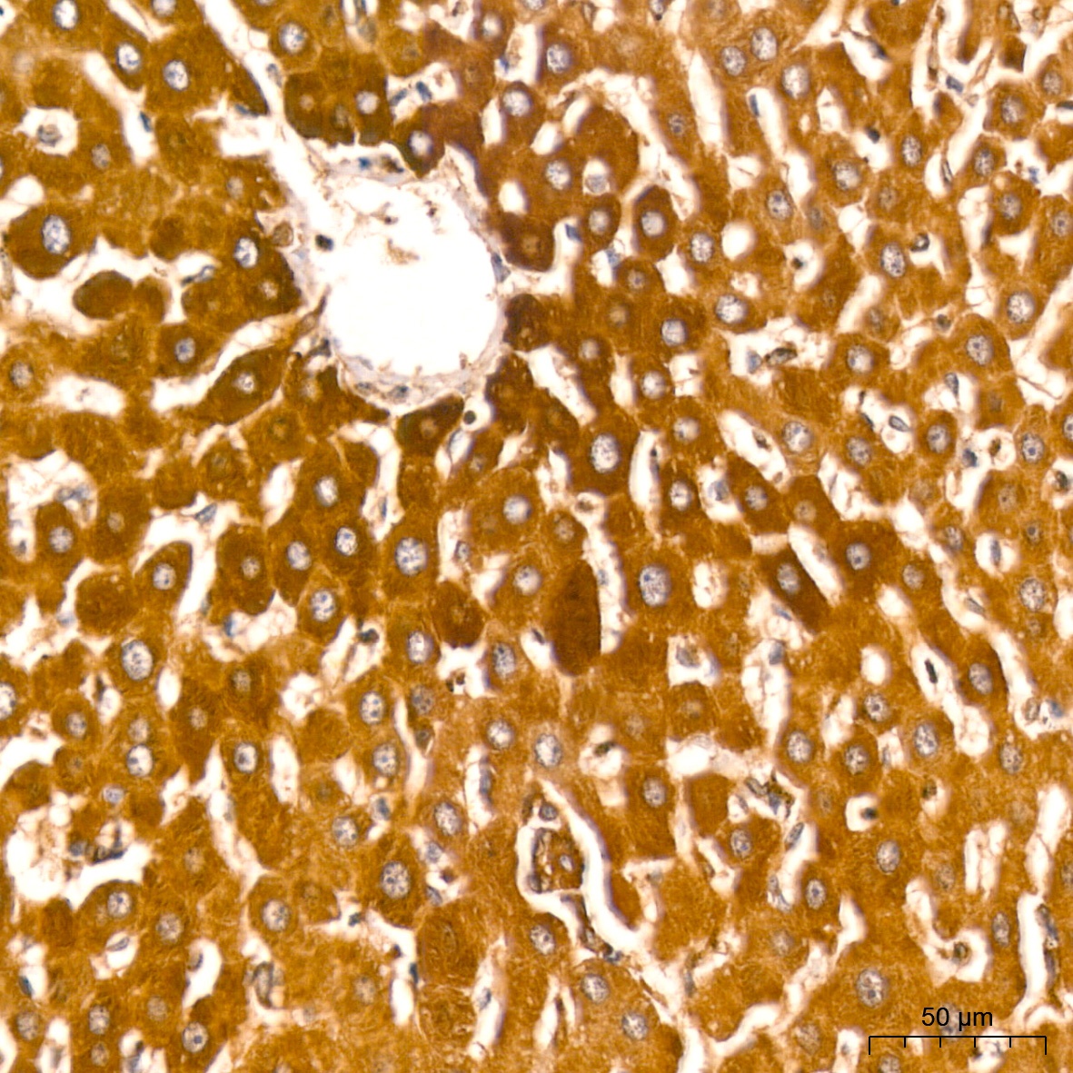
Immunohistochemistry analysis of paraffin-embedded Rat liver tissue using ATG3 Rabbit mAb (CAB19594) at a dilution of 1:200 (40x lens). High pressure antigen retrieval performed with 0.01M Tris-EDTA Buffer (pH 9.0) prior to IHC staining.
Immunohistochemistry analysis of paraffin-embedded Mouse kidney tissue using ATG3 Rabbit mAb (CAB19594) at a dilution of 1:200 (40x lens). High pressure antigen retrieval performed with 0.01M Citrate buffer (pH 6.0) prior to IHC staining.