The ATP13A2 Antibody (CAB13083) is a high-quality antibody developed for reliable detection and analysis of target proteins. The antibody, generated in rabbits, exhibits high reactivity with human samples and has been validated for use in various applications, including Western blot and immunohistochemistry.ATP13A2, also known as PARK9, functions as a lysosomal ATPase involved in maintaining lysosomal pH and metal ion homeostasis. Mutations in the ATP13A2 gene have been linked to lysosomal dysfunction and the pathogenesis of Parkinson's disease, highlighting its importance in neuronal health and survival.
This antibody is validated for use in WB, IF/ICC, ELISA applications and has demonstrated reactivity against Human, Mouse, Rat samples.
Product Name:
ATP13A2 Antibody
SKU:
CAB13083
Size:
20μL, 100μL
Reactivity:
Human, Mouse, Rat
Conjugate:
Unconjugated
Immunogen:
Recombinant protein (or fragment).This information is considered to be commercially sensitive.
Recommended starting concentration is 1 μg/mL. Please optimize the concentration based on your specific assay requirements.
Synonyms:
CLN12, KRPPD, PARK9, SPG78, HSA9947, ATP13A2
Positive Sample:
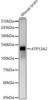
Mouse brain
Cellular Localization:
Lysosome, Membrane, Multi-Pass Membrane Protein.
Calculated MW:
129kDa
Observed MW:
130kDa
This gene encodes a member of the P5 subfamily of ATPases which transports inorganic cations as well as other substrates. Mutations in this gene are associated with Kufor-Rakeb syndrome (KRS), also referred to as Parkinson disease 9. Multiple transcript variants encoding different isoforms have been found for this gene.
Purification Method
Affinity purification
Gene ID
23400
RRID
AB_2759931
Buffer Information
Store at -20℃. Avoid freeze / thaw cycles. Buffer: PBS containing 50% glycerol, preserved with proclin300 or sodium azide, pH 7.3.
Western blot analysis of lysates from Mouse brain, using ATP13A2 Rabbit pAb (CAB13083) at 1:500 dilution. Secondary antibody: HRP-conjugated Goat anti-Rabbit IgG (H+L) (CABS014) at 1:10000 dilution. Lysates/proteins: 25μg per lane. Blocking buffer: 3% nonfat dry milk in TBST. Detection: ECL Basic Kit (AbGn00020). Exposure time: 60s.
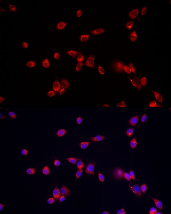
Immunofluorescence analysis of NIH/3T3 cells using ATP13A2 Rabbit pAb (CAB13083) at dilution of 1:50 (40x lens). Secondary antibody: Cy3-conjugated Goat anti-Rabbit IgG (H+L) (CABS007) at 1:500 dilution. Blue: DAPI for nuclear staining.