The ATPB Monoclonal Antibody (CAB11214) is a high-quality antibody developed for reliable detection and analysis of target proteins. This antibody, produced using rabbit-derived monoclonal technology, exhibits high specificity and sensitivity towards ATPB in a variety of experimental settings.ATPB, also known as ATP synthase subunit beta, plays a key role in ATP production through oxidative phosphorylation in the mitochondria. Dysregulation of ATPB expression or function can have profound effects on cellular energy metabolism, making it a crucial target for investigation in studies related to metabolism, aging, and various diseases.Validated for use in techniques such as immunohistochemistry and immunofluorescence, the ATPB Rabbit Monoclonal Antibody enables precise detection and visualization of ATPB localization within cells and tissues.
This antibody is validated for use in WB, IHC-P, IF/ICC, ELISA applications and has demonstrated reactivity against Human, Mouse, Rat samples.
Product Name:
ATPB Monoclonal Antibody
SKU:
CAB11214
Size:
20μL, 100μL
Reactivity:
Human, Mouse, Rat
Clone Number:
ARC53533
Conjugate:
Unconjugated
Immunogen:
Recombinant protein (or fragment).This information is considered to be commercially sensitive.
Recommended starting concentration is 1 μg/mL. Please optimize the concentration based on your specific assay requirements.
Synonyms:
ATP5B, ATPMB, ATPSB, HUMOP2, HEL-S-271, ATPB
Positive Sample:
HeLa, RAW 264.7, PC-12, Mouse heart
Cellular Localization:
Mitochondrion, Mitochondrion Inner Membrane.
Calculated MW:
57kDa
Observed MW:
57kDa
This gene encodes a subunit of mitochondrial ATP synthase. Mitochondrial ATP synthase catalyzes ATP synthesis, utilizing an electrochemical gradient of protons across the inner membrane during oxidative phosphorylation. ATP synthase is composed of two linked multi-subunit complexes: the soluble catalytic core, F1, and the membrane-spanning component, Fo, comprising the proton channel. The catalytic portion of mitochondrial ATP synthase consists of 5 different subunits (alpha, beta, gamma, delta, and epsilon) assembled with a stoichiometry of 3 alpha, 3 beta, and a single representative of the other 3. The proton channel consists of three main subunits (a, b, c). This gene encodes the beta subunit of the catalytic core.
Purification Method
Affinity purification
Gene ID
506
RRID
AB_2861523
Buffer Information
Store at -20℃. Avoid freeze / thaw cycles. Buffer: PBS containing 50% glycerol and 0.05% BSA, preserved with proclin300 or sodium azide, pH 7.3.
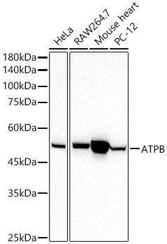
Western blot analysis of various lysates using ATPB Rabbit mAb (CAB11214) at1:50000 dilution. Secondary antibody: HRP-conjugated Goat anti-Rabbit IgG (H+L) (CABS014) at 1:10000 dilution. Lysates/proteins: 25μg per lane. Blocking buffer: 3% nonfat dry milk in TBST. Detection: ECL Basic Kit (AbGn00020). Exposure time: 10s.
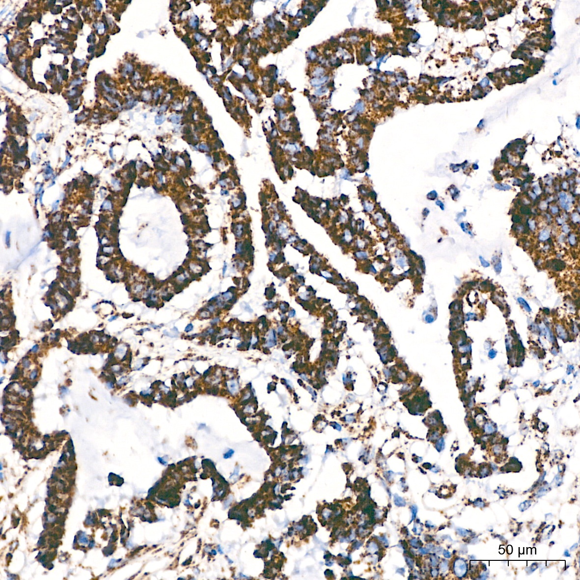
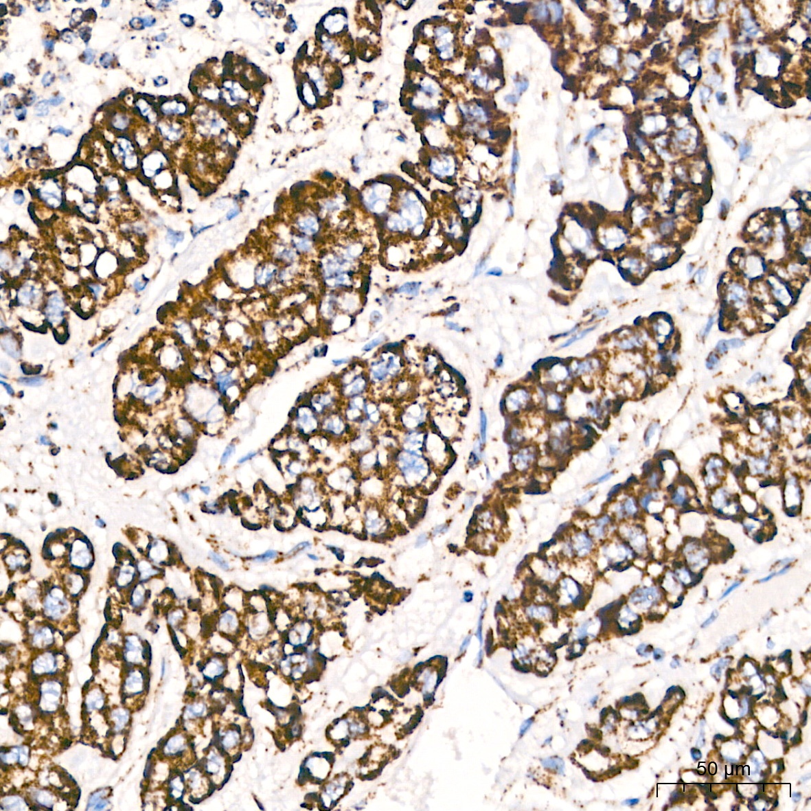
Immunohistochemistry analysis of paraffin-embedded Human colon carcinoma tissue using ATPB Rabbit mAb (CAB11214) at a dilution of 1:200 (40x lens). High pressure antigen retrieval performed with 0.01M Citrate Buffer (pH 6.0) prior to IHC staining.
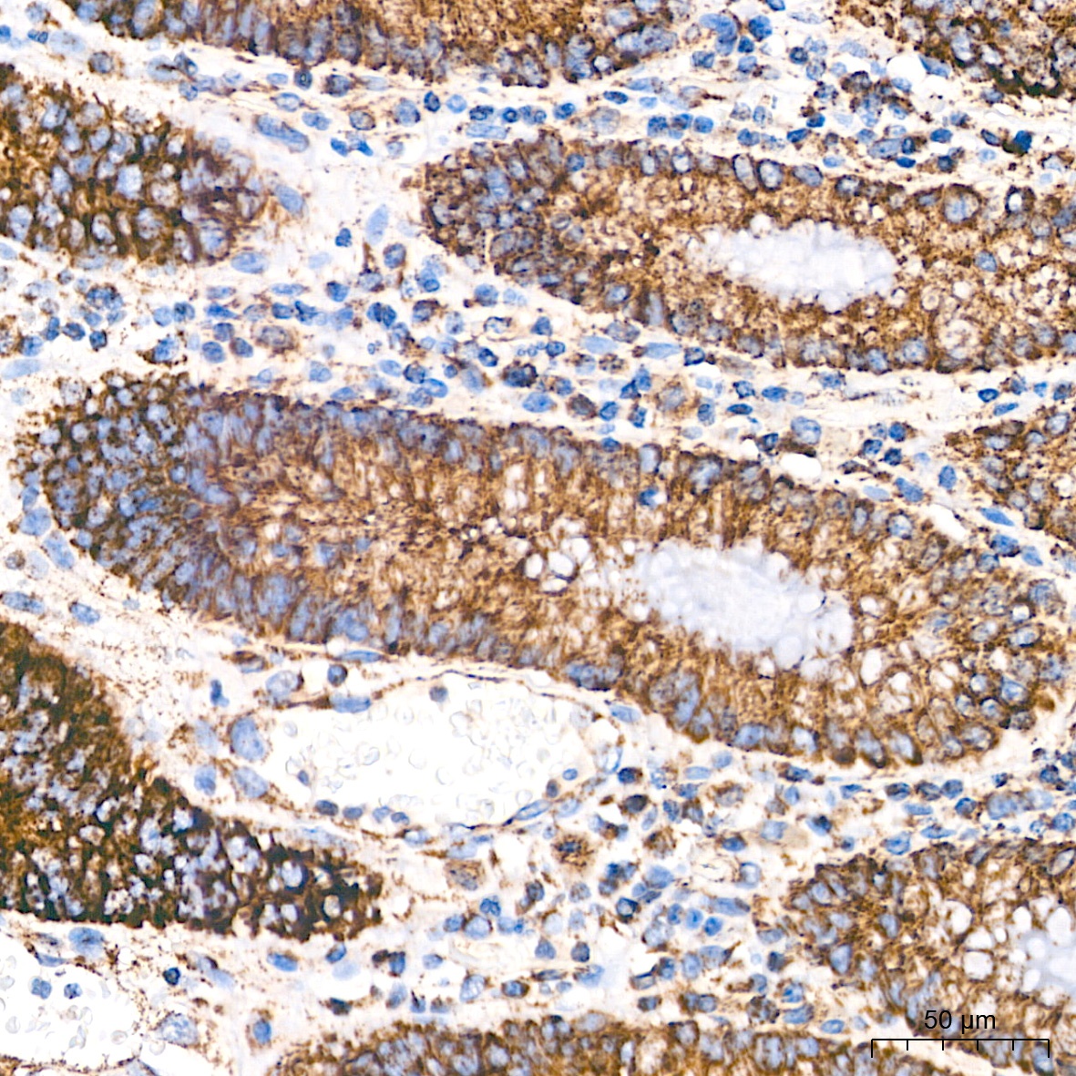
Immunohistochemistry analysis of paraffin-embedded Human colon tissue using ATPB Rabbit mAb (CAB11214) at a dilution of 1:200 (40x lens). High pressure antigen retrieval performed with 0.01M Citrate Buffer (pH 6.0) prior to IHC staining.
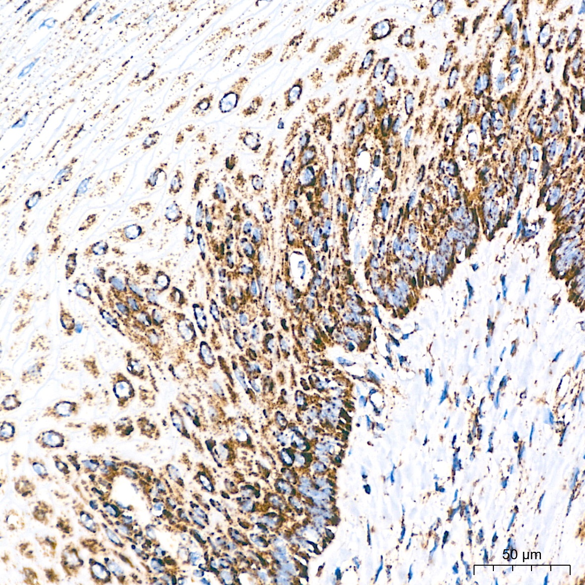
Immunohistochemistry analysis of paraffin-embedded Human esophagus tissue using ATPB Rabbit mAb (CAB11214) at a dilution of 1:200 (40x lens). High pressure antigen retrieval performed with 0.01M Citrate Buffer (pH 6.0) prior to IHC staining.
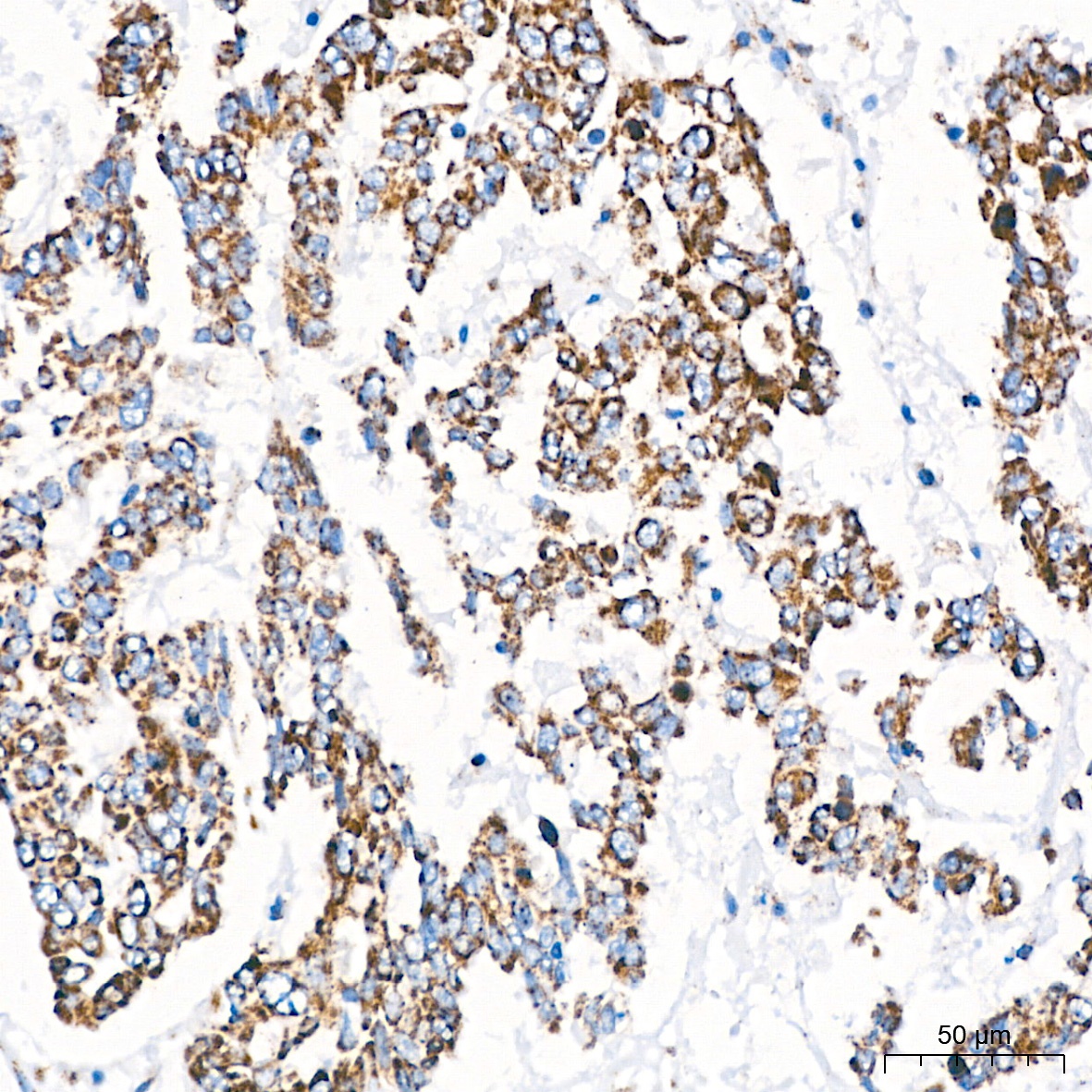
Immunohistochemistry analysis of paraffin-embedded Human liver cancer tissue using ATPB Rabbit mAb (CAB11214) at a dilution of 1:200 (40x lens). High pressure antigen retrieval performed with 0.01M Citrate Buffer (pH 6.0) prior to IHC staining.
Immunohistochemistry analysis of paraffin-embedded Human lung squamous carcinoma tissue using ATPB Rabbit mAb (CAB11214) at a dilution of 1:200 (40x lens). High pressure antigen retrieval performed with 0.01M Citrate Buffer (pH 6.0) prior to IHC staining.
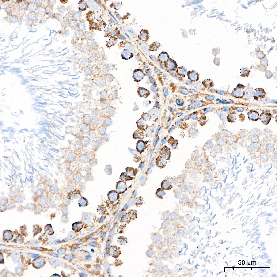
Immunohistochemistry analysis of paraffin-embedded Mouse testis tissue using ATPB Rabbit mAb (CAB11214) at a dilution of 1:200 (40x lens). High pressure antigen retrieval performed with 0.01M Citrate Buffer (pH 6.0) prior to IHC staining.
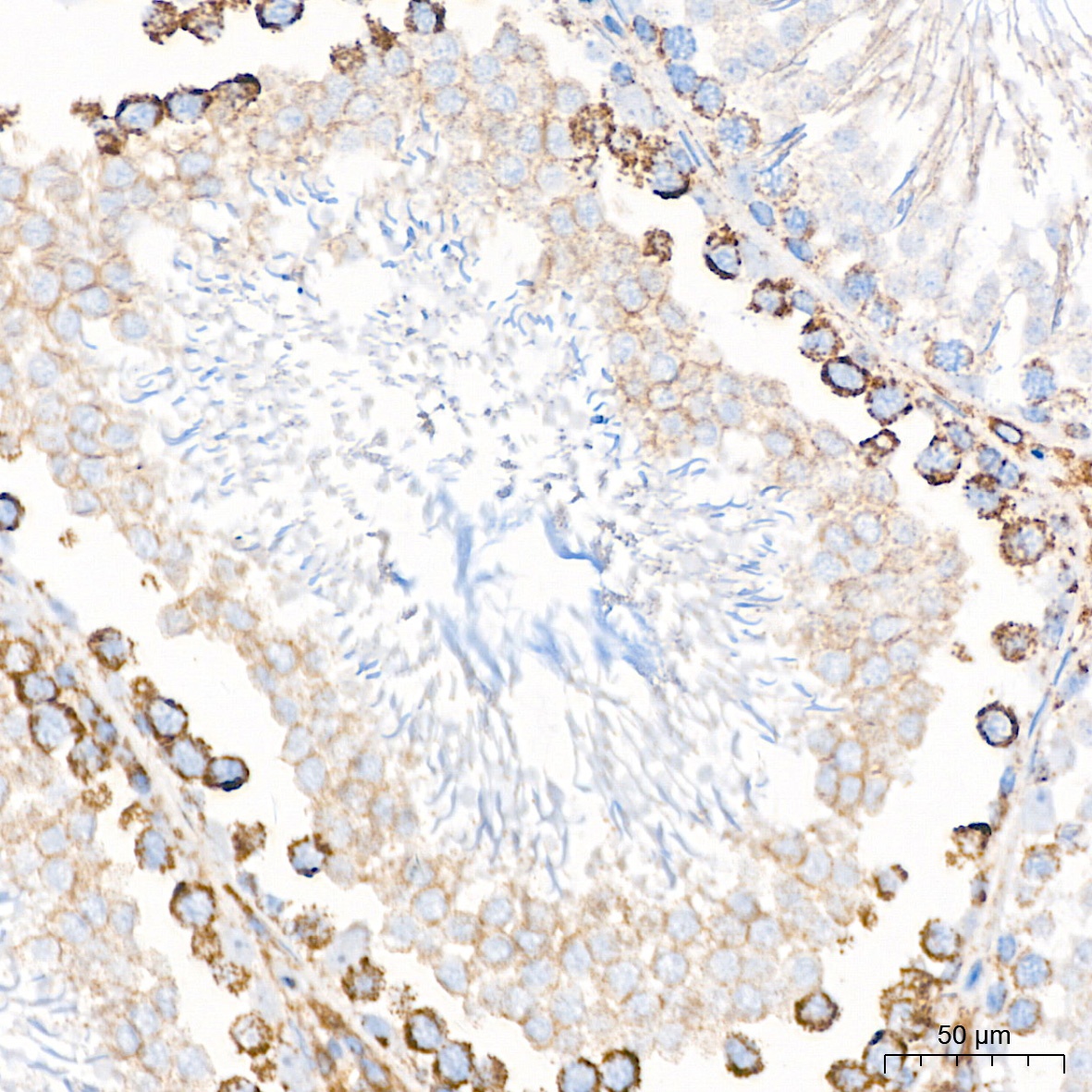
Immunohistochemistry analysis of paraffin-embedded Rat testis tissue using ATPB Rabbit mAb (CAB11214) at a dilution of 1:200 (40x lens). High pressure antigen retrieval performed with 0.01M Citrate Buffer (pH 6.0) prior to IHC staining.
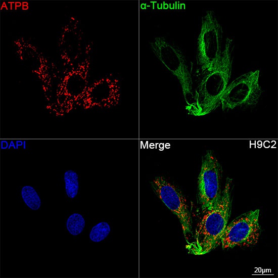
Confocal imaging of H9C2 cells using ATPB Rabbit mAb (CAB11214, dilution 1:200) followed by a further incubation with Cy3-conjugated Goat anti-Rabbit IgG (H+L) (CABS007, dilution 1:500) (Red). The cells were counterstained with α-Tubulin Mouse mAb (AC012, dilution 1:400) followed by incubation with ABflo® 488-conjugated Goat Anti-Mouse IgG (H+L) Ab (CABS076, dilution 1:500) (Green). DAPI was used for nuclear staining (Blue). Objective: 100x.
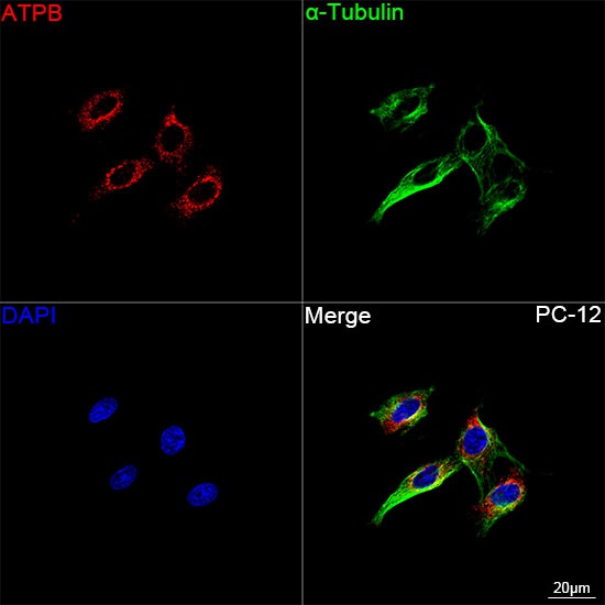
Confocal imaging of PC-12 cells using ATPB Rabbit mAb (CAB11214, dilution 1:200) followed by a further incubation with Cy3-conjugated Goat anti-Rabbit IgG (H+L) (CABS007, dilution 1:500) (Red). The cells were counterstained with α-Tubulin Mouse mAb (AC012, dilution 1:400) followed by incubation with ABflo® 488-conjugated Goat Anti-Mouse IgG (H+L) Ab (CABS076, dilution 1:500) (Green). DAPI was used for nuclear staining (Blue). Objective: 100x.
Confocal imaging of NIH/3T3 cells using ATPB Rabbit mAb (CAB11214, dilution 1:200) followed by a further incubation with Cy3 Goat Anti-Rabbit IgG (H+L) (CABS007, dilution 1:500) (Red). The cells were counterstained with α-Tubulin Mouse mAb (AC012, dilution 1:400) followed by incubation with ABflo® 488-conjugated Goat Anti-Mouse IgG (H+L) Ab (CABS076, dilution 1:500) (Green). DAPI was used for nuclear staining (Blue). Objective: 100x.