The ATP6 Antibody (CAB8193) is a high-quality antibody developed for reliable detection and analysis of target proteins. This antibody is produced through immunization of rabbits and exhibits high reactivity with human samples, making it suitable for use in Western blot applications. By targeting the ATP6 protein, this antibody enables researchers to detect and analyze ATP6 levels in various cell types, facilitating studies in bioenergetics, metabolism, and mitochondrial function.ATP6, a subunit of the ATP synthase complex, plays a crucial role in cellular energy production through oxidative phosphorylation. Dysregulation of ATP6 has been implicated in various diseases, including mitochondrial disorders, neurodegenerative diseases, and cancer.
This antibody is validated for use in WB, IHC-P, IF/ICC, ELISA applications and has demonstrated reactivity against Human, Mouse samples.
Product Name:
ATP6 Antibody
SKU:
CAB8193
Size:
20μL, 100μL
Reactivity:
Human, Mouse
Conjugate:
Unconjugated
Immunogen:
Synthetic peptide. This information is considered to be commercially sensitive.
Predicted to contribute to proton-transporting ATP synthase activity, rotational mechanism. Predicted to be involved in mitochondrial ATP synthesis coupled proton transport. Located in mitochondrion. Is expressed in several structures, including alimentary system; brain; heart; integumental system; and sensory organ. Human ortholog(s) of this gene implicated in Leber hereditary optic neuropathy; NARP syndrome; Parkinson's disease; multiple sclerosis; and systemic lupus erythematosus. Orthologous to human MT-ATP6 (mitochondrially encoded ATP synthase membrane subunit 6).
Purification Method
Affinity purification
Gene ID
17705
RRID
AB_2768510
Buffer Information
Store at -20℃. Avoid freeze / thaw cycles. Buffer: PBS containing 50% glycerol, preserved with proclin300 or sodium azide, pH 7.3.
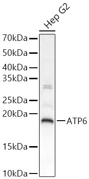
Western blot analysis of lysates from Hep G2 cells, using ATP6 Rabbit pAb (CAB8193) at 1:2000 dilution. Secondary antibody: HRP-conjugated Goat anti-Rabbit IgG (H+L) (CABS014) at 1:10000 dilution. Lysates/proteins: 25 μg per lane. Blocking buffer: 3% nonfat dry milk in TBST. Detection: ECL Basic Kit (AbGn00020). Exposure time: 20 s.
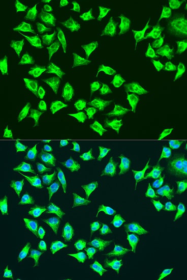
Immunofluorescence analysis of U-2 OS cells using ATP6 Rabbit pAb (CAB8193) at dilution of 1:100. Secondary antibody: Cy3-conjugated Goat anti-Rabbit IgG (H+L) (CABS007) at 1:500 dilution. Blue: DAPI for nuclear staining.