The AURKA Antibody (CAB2121) is a high-quality antibody developed for reliable detection and analysis of target proteins. This polyclonal antibody, raised in rabbits, exhibits high reactivity with human samples and is validated for use in various applications, including Western blotting.Aurora Kinase A is a crucial player in cell division, functioning to ensure accurate chromosome segregation and proper cell division. Dysregulation of Aurora Kinase A has been linked to cancer development and progression, making it an attractive target for cancer research and therapy development.
This antibody is validated for use in WB, IF/ICC, ELISA applications and has demonstrated reactivity against Human, Mouse samples.
Product Name:
AURKA Antibody
SKU:
CAB2121
Size:
20μL, 100μL
Reactivity:
Human, Mouse
Conjugate:
Unconjugated
Immunogen:
Synthetic peptide. This information is considered to be commercially sensitive.
Recommended starting concentration is 1 μg/mL. Please optimize the concentration based on your specific assay requirements.
Synonyms:
AIK, ARK1, AURA, BTAK, STK6, STK7, STK15, PPP1R47, AURKA
Positive Sample:
HeLa
Cellular Localization:
Cytoplasm, Centriole, Centrosome, Cilium Basal Body, Cytoskeleton, Microtubule Organizing Center, Spindle Pole.
Calculated MW:
46kDa
Observed MW:
46kDa
The protein encoded by this gene is a cell cycle-regulated kinase that appears to be involved in microtubule formation and/or stabilization at the spindle pole during chromosome segregation. The encoded protein is found at the centrosome in interphase cells and at the spindle poles in mitosis. This gene may play a role in tumor development and progression. A processed pseudogene of this gene has been found on chromosome 1, and an unprocessed pseudogene has been found on chromosome 10. Multiple transcript variants encoding the same protein have been found for this gene.
Purification Method
Affinity purification
Gene ID
6790
RRID
AB_2764140
Buffer Information
Store at -20℃. Avoid freeze / thaw cycles. Buffer: PBS containing 50% glycerol, preserved with proclin300 or sodium azide, pH 7.3.
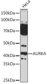
Western blot analysis of lysates from HeLa cells, using AURKA Rabbit pAb (CAB2121) at 1:500 dilution. Secondary antibody: HRP-conjugated Goat anti-Rabbit IgG (H+L) (CABS014) at 1:10000 dilution. Lysates/proteins: 25μg per lane. Blocking buffer: 3% nonfat dry milk in TBST. Detection: ECL Basic Kit (AbGn00020). Exposure time: 90s.
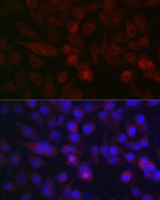
Immunofluorescence analysis of HeLa cells using AURKA Rabbit pAb (CAB2121) at dilution of 1:100 (40x lens). Secondary antibody: Cy3-conjugated Goat anti-Rabbit IgG (H+L) (CABS007) at 1:500 dilution. Blue: DAPI for nuclear staining.